6M2I | pdb_00006m2i

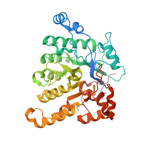
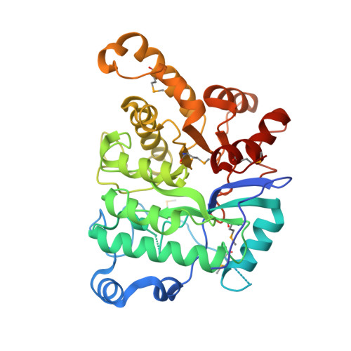
Structure of the 2-Aminoisobutyric acid Monooxygenase Hydroxylase


Experimental Data Snapshot

  • Method: X-RAY DIFFRACTION
  • Resolution: 2.45 Å
  • R-Value Free: 
    0.236 (Depositor), 0.240 (DCC) 
  • R-Value Work: 
    0.184 (Depositor), 0.186 (DCC) 
  • R-Value Observed: 
    0.187 (Depositor) 

Starting Model: experimental
View more details

wwPDB Validation 3D Report Full Report

Validation slider image for 6M2I

Ligand Structure Quality Assessment 


This is version 1.4 of the entry. See complete history. 

Literature

A three-component monooxygenase from Rhodococcus wratislaviensis may expand industrial applications of bacterial enzymes.

Hibi, M., Fukuda, D., Kenchu, C., Nojiri, M., Hara, R., Takeuchi, M., Aburaya, S., Aoki, W., Mizutani, K., Yasohara, Y., Ueda, M., Mikami, B., Takahashi, S., Ogawa, J.

(2021) Commun Biol 4: 16-16

  • DOI: https://doi.org/10.1038/s42003-020-01555-3
  • Primary Citation Related Structures: 
    6M1W, 6M2I

  • PubMed Abstract: 

    The high-valent iron-oxo species formed in the non-heme diiron enzymes have high oxidative reactivity and catalyze difficult chemical reactions. Although the hydroxylation of inert methyl groups is an industrially promising reaction, utilizing non-heme diiron enzymes as such a biocatalyst has been difficult. Here we show a three-component monooxygenase system for the selective terminal hydroxylation of α-aminoisobutyric acid (Aib) into α-methyl-D-serine. It consists of the hydroxylase component, AibH1H2, and the electron transfer component. Aib hydroxylation is the initial step of Aib catabolism in Rhodococcus wratislaviensis C31-06, which has been fully elucidated through a proteome analysis. The crystal structure analysis revealed that AibH1H2 forms a heterotetramer of two amidohydrolase superfamily proteins, of which AibHm2 is a non-heme diiron protein and functions as a catalytic subunit. The Aib monooxygenase was demonstrated to be a promising biocatalyst that is suitable for bioprocesses in which the inert C-H bond in methyl groups need to be activated.


  • Organizational Affiliation: 
    • Biotechnology Research Center and Department of Biotechnology, Toyama Prefectural University, 5180 Kurokawa, Imizu, Toyama, 939-0398, Japan. hibi@pu-toyama.ac.jp.

Macromolecule Content 

  • Total Structure Weight: 87.79 kDa 
  • Atom Count: 5,863 
  • Modeled Residue Count: 708 
  • Deposited Residue Count: 763 
  • Unique protein chains: 2

Macromolecules

Find similar proteins by:|  3D Structure
Entity ID: 1
MoleculeChains  Sequence LengthOrganismDetailsImage
Amidohydrolase385Rhodococcus wratislaviensisMutation(s): 0 
Gene Names: Rhow_000804
UniProt
Find proteins for A0A402C2V4 (Rhodococcus wratislaviensis)
Explore A0A402C2V4 
Go to UniProtKB:  A0A402C2V4
Entity Groups
Sequence Clusters30% Identity50% Identity70% Identity90% Identity95% Identity100% Identity
UniProt GroupA0A402C2V4
Sequence Annotations
Expand
Reference Sequence
Find similar proteins by:|  3D Structure
Entity ID: 2
MoleculeChains  Sequence LengthOrganismDetailsImage
Amidohydrolase378Rhodococcus wratislaviensisMutation(s): 0 
Gene Names: Rhow_000803
UniProt
Find proteins for A0A402C2Q3 (Rhodococcus wratislaviensis)
Explore A0A402C2Q3 
Go to UniProtKB:  A0A402C2Q3
Entity Groups
Sequence Clusters30% Identity50% Identity70% Identity90% Identity95% Identity100% Identity
UniProt GroupA0A402C2Q3
Sequence Annotations
Expand
Reference Sequence

Small Molecules

Ligands 4 Unique
IDChains Name / Formula / InChI Key2D Diagram3D Interactions
SO4

Query on SO4


Download:Ideal Coordinates CCD File
D [auth A],
E [auth A],
F [auth A],
G [auth A],
T [auth B]
SULFATE ION
O4 S
QAOWNCQODCNURD-UHFFFAOYSA-L
ZN

Query on ZN


Download:Ideal Coordinates CCD File
C [auth A]ZINC ION
Zn
PTFCDOFLOPIGGS-UHFFFAOYSA-N
EDO
(Subject of Investigation/LOI)

Query on EDO


Download:Ideal Coordinates CCD File
AA [auth B]
H [auth A]
I [auth A]
J [auth A]
K [auth A]
AA [auth B],
H [auth A],
I [auth A],
J [auth A],
K [auth A],
L [auth A],
M [auth A],
N [auth A],
O [auth A],
P [auth A],
Q [auth A],
U [auth B],
V [auth B],
W [auth B],
X [auth B],
Y [auth B],
Z [auth B]
1,2-ETHANEDIOL
C2 H6 O2
LYCAIKOWRPUZTN-UHFFFAOYSA-N
FE

Query on FE


Download:Ideal Coordinates CCD File
R [auth B],
S [auth B]
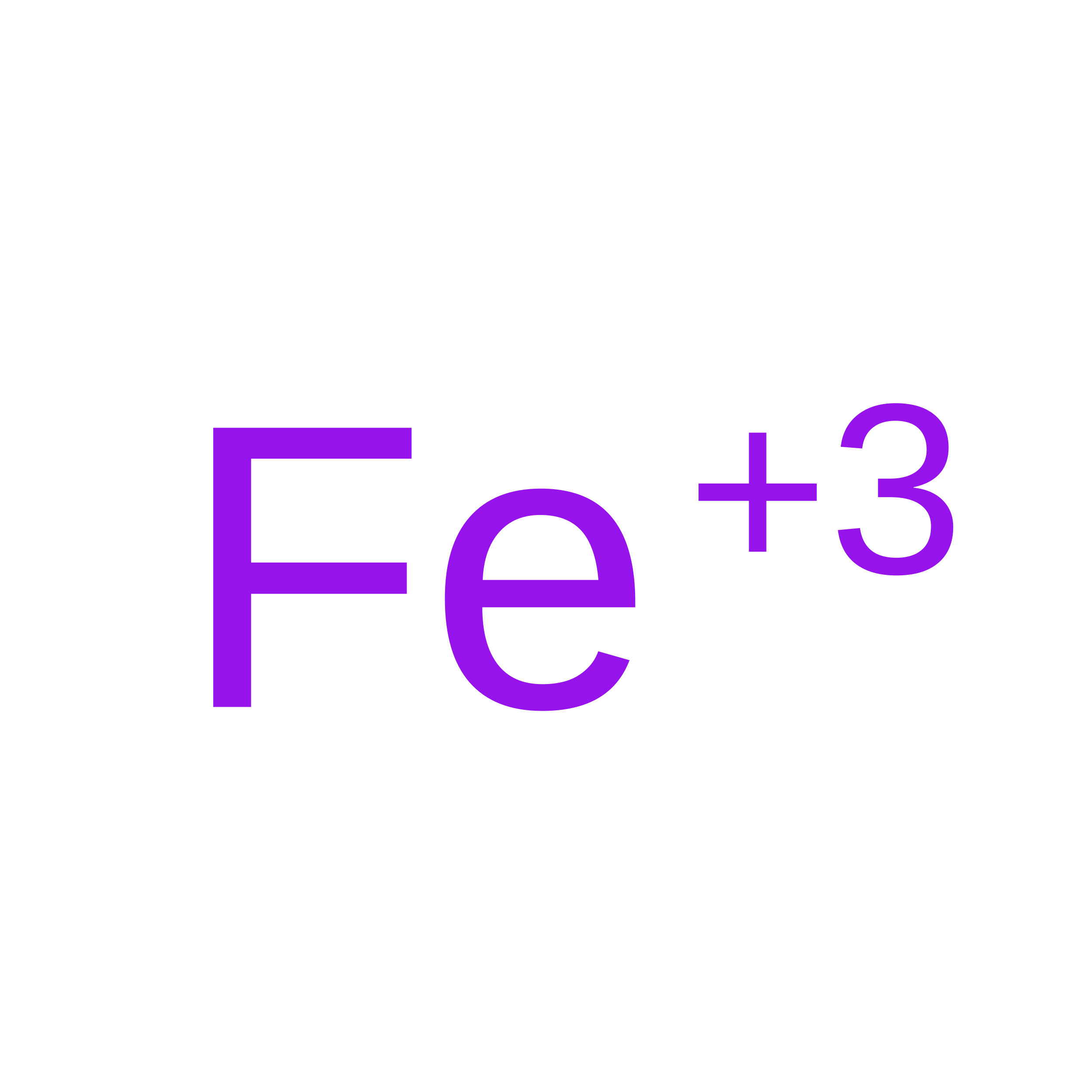
FE (III) ION
Fe
VTLYFUHAOXGGBS-UHFFFAOYSA-N
Modified Residues  1 Unique
IDChains TypeFormula2D DiagramParent
MSE
Query on MSE
A
L-PEPTIDE LINKINGC5 H11 N O2 SeMET

Experimental Data & Validation

Experimental Data

  • Method: X-RAY DIFFRACTION
  • Resolution: 2.45 Å
  • R-Value Free:  0.236 (Depositor), 0.240 (DCC) 
  • R-Value Work:  0.184 (Depositor), 0.186 (DCC) 
  • R-Value Observed: 0.187 (Depositor) 
Space Group: C 2 2 21
Unit Cell:
Length ( Å )Angle ( ˚ )
a = 123.751α = 90
b = 206.684β = 90
c = 91.489γ = 90
Software Package:
Software NamePurpose
PHENIXrefinement
XDSdata reduction
XDSdata scaling
PHENIXphasing

Structure Validation

View Full Validation Report


Ligand Structure Quality Assessment 


Entry History 

Deposition Data

Revision History  (Full details and data files)

  • Version 1.0: 2021-01-06
    Type: Initial release
  • Version 1.1: 2021-01-27
    Changes: Database references
  • Version 1.2: 2023-11-29
    Changes: Data collection, Database references, Refinement description
  • Version 1.3: 2024-11-13
    Changes: Structure summary
  • Version 1.4: 2025-03-12
    Changes: Database references