6M0E | pdb_00006m0e

Beijerinckia indica beta-fructosyltransferase complexed with fructose


Experimental Data Snapshot

  • Method: X-RAY DIFFRACTION
  • Resolution: 1.35 Å
  • R-Value Free: 
    0.166 (Depositor), 0.175 (DCC) 
  • R-Value Work: 
    0.150 (Depositor) 

Starting Model: experimental
View more details

wwPDB Validation 3D Report Full Report

Validation slider image for 6M0E

Ligand Structure Quality Assessment 


This is version 1.5 of the entry. See complete history

Literature

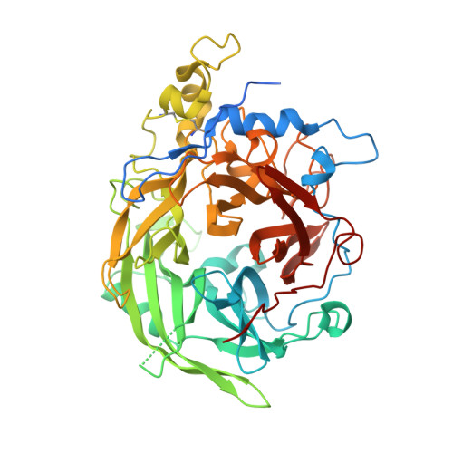
Crystal structure of a glycoside hydrolase family 68 beta-fructosyltransferase from Beijerinckia indica subsp. indica in complex with fructose.

Tonozuka, T.Kitamura, J.Nagaya, M.Kawai, R.Nishikawa, A.Hirano, K.Tamura, K.Fujii, T.Tochio, T.

(2020) Biosci Biotechnol Biochem 84: 2508-2520

  • DOI: https://doi.org/10.1080/09168451.2020.1804317
  • Primary Citation Related Structures: 
    6M0D, 6M0E

  • PubMed Abstract: 

    An enzyme belonging to glycoside hydrolase family 68 (GH68) from Beijerinckia indica subsp. indica NBRC 3744 was expressed in Escherichia coli . Biochemical characterization showed that the enzyme was identified to be a β-fructosyltransferase (BiBftA). Crystallization of a full-length BiBftA was initially attempted, but no crystals were obtained. We constructed a variant in which 5 residues (Pro199-Gly203) and 13 residues (Leu522-Gln534) in potentially flexible regions were deleted, and we successfully crystallized this variant BiBftA. BiBftA is composed of a five-bladed β-propeller fold as in other GH68 enzymes. The structure of BiBftA in complex with fructose unexpectedly indicated that one β-fructofuranose (β-Fru f ) molecule and one β-fructopyranose molecule bind to the catalytic pocket. The orientation of β-Fru f at subsite -1 is tilted from the orientation observed in most GH68 enzymes, presenting a second structure of a GH68 enzyme in complex with the tilted binding mode of β-Fru f.


  • Organizational Affiliation
    • Department of Applied Biological Science, Tokyo University of Agriculture and Technology , Tokyo, Japan.

Macromolecule Content 

  • Total Structure Weight: 57.98 kDa 
  • Atom Count: 4,350 
  • Modeled Residue Count: 477 
  • Deposited Residue Count: 523 
  • Unique protein chains: 1

Macromolecules

Find similar proteins by:|  3D Structure
Entity ID: 1
MoleculeChains  Sequence LengthOrganismDetailsImage
Levansucrase523Beijerinckia indica subsp. indica ATCC 9039Mutation(s): 0 
Gene Names: Bind_2021
EC: 2.4.1.10
UniProt
Find proteins for B2IF78 (Beijerinckia indica subsp. indica (strain ATCC 9039 / DSM 1715 / NCIMB 8712))
Explore B2IF78 
Go to UniProtKB:  B2IF78
Entity Groups
Sequence Clusters30% Identity50% Identity70% Identity90% Identity95% Identity100% Identity
UniProt GroupB2IF78
Sequence Annotations
Expand
Reference Sequence

Small Molecules

Ligands 3 Unique
IDChains Name / Formula / InChI Key2D Diagram3D Interactions
BDF
(Subject of Investigation/LOI)

Query on BDF


Download:Ideal Coordinates CCD File
F [auth A],
G [auth A],
H [auth A],
I [auth A]
beta-D-fructopyranose
C6 H12 O6
LKDRXBCSQODPBY-ARQDHWQXSA-N
FRU
(Subject of Investigation/LOI)

Query on FRU


Download:Ideal Coordinates CCD File
E [auth A]beta-D-fructofuranose
C6 H12 O6
RFSUNEUAIZKAJO-ARQDHWQXSA-N
MG
(Subject of Investigation/LOI)

Query on MG


Download:Ideal Coordinates CCD File
B [auth A],
C [auth A],
D [auth A]
MAGNESIUM ION
Mg
JLVVSXFLKOJNIY-UHFFFAOYSA-N

Experimental Data & Validation

Experimental Data

  • Method: X-RAY DIFFRACTION
  • Resolution: 1.35 Å
  • R-Value Free:  0.166 (Depositor), 0.175 (DCC) 
  • R-Value Work:  0.150 (Depositor) 
Space Group: P 21 21 21
Unit Cell:
Length ( Å )Angle ( ˚ )
a = 54.377α = 90
b = 81.521β = 90
c = 99.081γ = 90
Software Package:
Software NamePurpose
REFMACrefinement
XDSdata reduction
SCALAdata scaling
MOLREPphasing

Structure Validation

View Full Validation Report


Ligand Structure Quality Assessment 


Entry History 

Deposition Data

  • Released Date: 2020-08-12 
  • Deposition Author(s): Tonozuka, T.

Revision History  (Full details and data files)

  • Version 1.0: 2020-08-12
    Type: Initial release
  • Version 1.1: 2020-08-19
    Changes: Database references
  • Version 1.2: 2020-10-14
    Changes: Database references
  • Version 1.3: 2020-11-18
    Changes: Database references
  • Version 1.4: 2023-11-29
    Changes: Data collection, Database references, Derived calculations, Refinement description
  • Version 1.5: 2024-11-06
    Changes: Structure summary