Aromatic Interactions Drive the Coupled Folding and Binding of the Intrinsically Disordered Sesbania mosaic Virus VPg Protein.
Dixit, K., Karanth, N.M., Nair, S., Kumari, K., Chakrabarti, K.S., Savithri, H.S., Sarma, S.P.(2020) Biochemistry 59: 4663-4680
- PubMed: 33269926 Search on PubMed
- DOI: https://doi.org/10.1021/acs.biochem.0c00721
- Primary Citation Related Structures:
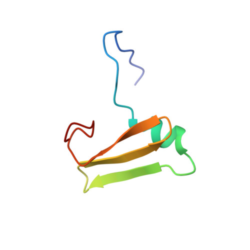
6LXF, 6M78 - PubMed Abstract:
The plant Sesbania mosaic virus [a (+)-ssRNA sobemovirus] VPg protein is intrinsically disordered in solution. For the virus life cycle, the VPg protein is essential for replication and for polyprotein processing that is carried out by a virus-encoded protease. The nuclear magnetic resonance (NMR)-derived tertiary structure of the protease-bound VPg shows it to have a novel tertiary structure with an α-β-β-β topology. The quaternary structure of the high-affinity protease-VPg complex (≈27 kDa) has been determined using HADDOCK protocols with NMR (residual dipolar coupling, dihedral angle, and nuclear Overhauser enhancement) restraints and mutagenesis data as inputs. The geometry of the complex is in excellent agreement with long-range orientational restraints such as residual dipolar couplings and ring-current shifts. A "vein" of aromatic residues on the protease surface is pivotal for the folding of VPg via intermolecular edge-to-face π···π stacking between Trp 271 and Trp 368 of the protease and VPg, respectively, and for the CH···π interactions between Leu 361 of VPg and Trp 271 of the protease. The structure of the protease-VPg complex provides a molecular framework for predicting sites of important posttranslational modifications such as RNA linkage and phosphorylation and a better understanding of the coupled folding upon binding of intrinsically disordered proteins. The structural data presented here augment the limited structural data available on viral proteins, given their propensity for structural disorder.
- Molecular Biophysics Unit, Indian Institute of Science, Bangalore, Karnataka 560012, India.
Organizational Affiliation: