6LVX | pdb_00006lvx

Crystal structure of TLR7/Cpd-1 (SM-374527) complex


Experimental Data Snapshot

  • Method: X-RAY DIFFRACTION
  • Resolution: 2.77 Å
  • R-Value Free: 
    0.252 (Depositor), 0.255 (DCC) 
  • R-Value Work: 
    0.212 (Depositor), 0.216 (DCC) 
  • R-Value Observed: 
    0.214 (Depositor) 

Starting Model: experimental
View more details

wwPDB Validation 3D Report Full Report

Validation slider image for 6LVX

Ligand Structure Quality Assessment 


This is version 1.2 of the entry. See complete history

Literature

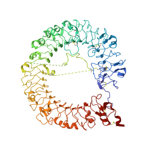
Structural analysis reveals TLR7 dynamics underlying antagonism.

Tojo, S.Zhang, Z.Matsui, H.Tahara, M.Ikeguchi, M.Kochi, M.Kamada, M.Shigematsu, H.Tsutsumi, A.Adachi, N.Shibata, T.Yamamoto, M.Kikkawa, M.Senda, T.Isobe, Y.Ohto, U.Shimizu, T.

(2020) Nat Commun 11: 5204-5204

  • DOI: https://doi.org/10.1038/s41467-020-19025-z
  • Primary Citation Related Structures: 
    6LVX, 6LVY, 6LVZ, 6LW0, 6LW1

  • PubMed Abstract: 

    Toll-like receptor 7 (TLR7) recognizes both microbial and endogenous RNAs and nucleosides. Aberrant activation of TLR7 has been implicated in several autoimmune diseases including systemic lupus erythematosus (SLE). Here, by modifying potent TLR7 agonists, we develop a series of TLR7-specific antagonists as promising therapeutic agents for SLE. These compounds protect mice against lethal autoimmunity. Combining crystallography and cryo-electron microscopy, we identify the open conformation of the receptor and reveal the structural equilibrium between open and closed conformations that underlies TLR7 antagonism, as well as the detailed mechanism by which TLR7-specific antagonists bind to their binding pocket in TLR7. Our work provides small-molecule TLR7-specific antagonists and suggests the TLR7-targeting strategy for treating autoimmune diseases.


  • Organizational Affiliation
    • Sumitomo Dainippon Pharma Co., Ltd., 3-1-98 Kasugade-naka, Konohana-ku, Osaka, 554-0022, Japan. shingo-tojo@ds-pharma.co.jp.

Macromolecule Content 

  • Total Structure Weight: 193.55 kDa 
  • Atom Count: 12,774 
  • Modeled Residue Count: 1,542 
  • Deposited Residue Count: 1,646 
  • Unique protein chains: 1

Macromolecules

Find similar proteins by:|  3D Structure
Entity ID: 1
MoleculeChains  Sequence LengthOrganismDetailsImage
Toll-like receptor 7
A, B
823Macaca mulattaMutation(s): 10 
Gene Names: TLR7
UniProt
Find proteins for B3Y653 (Macaca mulatta)
Explore B3Y653 
Go to UniProtKB:  B3Y653
Entity Groups
Sequence Clusters30% Identity50% Identity70% Identity90% Identity95% Identity100% Identity
UniProt GroupB3Y653
Glycosylation
Glycosylation Sites: 5
Sequence Annotations
Expand
Reference Sequence

Oligosaccharides

Help  
Entity ID: 2
MoleculeChains Length2D Diagram GlycosylationD Interactions
2-acetamido-2-deoxy-beta-D-glucopyranose-(1-4)-2-acetamido-2-deoxy-beta-D-glucopyranose
C
2N-Glycosylation
Glycosylation Resources
GlyTouCan: G42666HT
GlyCosmos: G42666HT
GlyGen: G42666HT

Small Molecules

Ligands 3 Unique
IDChains Name / Formula / InChI Key2D Diagram3D Interactions
EWL
(Subject of Investigation/LOI)

Query on EWL


Download:Ideal Coordinates CCD File
D [auth A],
P [auth B]
6-azanyl-2-butoxy-9-(phenylmethyl)-7H-purin-8-one
C16 H19 N5 O2
HEKGNUXGYUZNHH-UHFFFAOYSA-N
NAG

Query on NAG


Download:Ideal Coordinates CCD File
E [auth A]
F [auth A]
G [auth A]
H [auth A]
I [auth A]
E [auth A],
F [auth A],
G [auth A],
H [auth A],
I [auth A],
J [auth A],
K [auth A],
Q [auth B],
R [auth B],
S [auth B],
T [auth B]
2-acetamido-2-deoxy-beta-D-glucopyranose
C8 H15 N O6
OVRNDRQMDRJTHS-FMDGEEDCSA-N
SO4

Query on SO4


Download:Ideal Coordinates CCD File
L [auth A]
M [auth A]
N [auth A]
O [auth A]
U [auth B]
L [auth A],
M [auth A],
N [auth A],
O [auth A],
U [auth B],
V [auth B]
SULFATE ION
O4 S
QAOWNCQODCNURD-UHFFFAOYSA-L
Binding Affinity Annotations 
IDSourceBinding Affinity
EWL BindingDB:  6LVX EC50: 63.7 (nM) from 1 assay(s)

Experimental Data & Validation

Experimental Data

  • Method: X-RAY DIFFRACTION
  • Resolution: 2.77 Å
  • R-Value Free:  0.252 (Depositor), 0.255 (DCC) 
  • R-Value Work:  0.212 (Depositor), 0.216 (DCC) 
  • R-Value Observed: 0.214 (Depositor) 
Space Group: P 21 21 21
Unit Cell:
Length ( Å )Angle ( ˚ )
a = 98.754α = 90
b = 138.504β = 90
c = 148.462γ = 90
Software Package:
Software NamePurpose
REFMACrefinement
PDB_EXTRACTdata extraction
XDSdata reduction
Aimlessdata scaling
MOLREPphasing

Structure Validation

View Full Validation Report


Ligand Structure Quality Assessment 


Entry History 

Deposition Data

Revision History  (Full details and data files)

  • Version 1.0: 2020-11-11
    Type: Initial release
  • Version 1.1: 2023-11-29
    Changes: Data collection, Database references, Refinement description
  • Version 1.2: 2024-10-23
    Changes: Structure summary