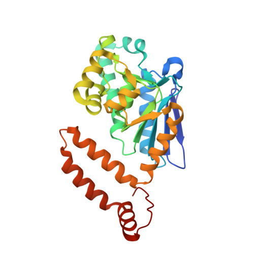
Structural and sequence comparisons of bacterial enoyl-CoA isomerase and enoyl-CoA hydratase.
Hwang, J., Jeong, C.S., Lee, C.W., Shin, S.C., Kim, H.W., Lee, S.G., Youn, U.J., Lee, C.S., Oh, T.J., Kim, H.J., Park, H., Park, H.H., Lee, J.H.(2020) J Microbiol 58: 606-613
- PubMed: 32323197 Search on PubMed
- DOI: https://doi.org/10.1007/s12275-020-0089-1
- Primary Citation Related Structures:
6LVO, 6LVP - PubMed Abstract:
Crystal structures of enoyl-coenzyme A (CoA) isomerase from Bosea sp. PAMC 26642 (BoECI) and enoyl-CoA hydratase from Hymenobacter sp. PAMC 26628 (HyECH) were determined at 2.35 and 2.70 Å resolution, respectively. BoECI and HyECH are members of the crotonase superfamily and are enzymes known to be involved in fatty acid degradation. Structurally, these enzymes are highly similar except for the orientation of their C-terminal helix domain. Analytical ultracentrifugation was performed to determine the oligomerization states of BoECI and HyECH revealing they exist as trimers in solution. However, their putative ligand-binding sites and active site residue compositions are dissimilar. Comparative sequence and structural analysis revealed that the active site of BoECI had one glutamate residue (Glu135), this site is occupied by an aspartate in some ECIs, and the active sites of HyECH had two highly conserved glutamate residues (Glu118 and Glu138). Moreover, HyECH possesses a salt bridge interaction between Glu98 and Arg152 near the active site. This interaction may allow the catalytic Glu118 residue to have a specific conformation for the ECH enzyme reaction. This salt bridge interaction is highly conserved in known bacterial ECH structures and ECI enzymes do not have this type of interaction. Collectively, our comparative sequential and structural studies have provided useful information to distinguish and classify two similar bacterial crotonase superfamily enzymes.
- Unit of Research for Practical Application, Korea Polar Research Institute, Incheon, 21990, Republic of Korea.
Organizational Affiliation: