6LL9 | pdb_00006ll9

Crystal structure of D-alanine-D-alanine ligase from Aeromonas hydrophila


Experimental Data Snapshot

  • Method: X-RAY DIFFRACTION
  • Resolution: 2.70 Å
  • R-Value Free: 
    0.350 (Depositor), 0.371 (DCC) 
  • R-Value Work: 
    0.295 (Depositor), 0.322 (DCC) 
  • R-Value Observed: 
    0.297 (Depositor) 

Starting Model: experimental
View more details

wwPDB Validation 3D Report Full Report

Validation slider image for 6LL9

This is version 1.2 of the entry. See complete history

Literature

Insights into the Inhibition ofAeromonas hydrophilad-Alanine-d-Alanine Ligase by Integration of Kinetics and Structural Analysis.

Zhang, Y.Gong, S.Wang, X.Muhammad, M.Li, Y.Meng, S.Li, Q.Liu, D.Zhang, H.

(2020) J Agric Food Chem 68: 7509-7519

  • DOI: https://doi.org/10.1021/acs.jafc.0c00682
  • Primary Citation Related Structures: 
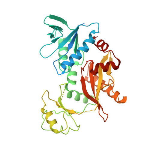
    6LL9

  • PubMed Abstract: 

    Aeromonas hydrophila , a pathogenic bacterium, is harmful to humans, domestic animals, and fishes and, moreover, of public health concern due to the emergence of multiple drug-resistant strains. The cell wall has been discovered as a novel and efficient drug target against bacteria, and d-alanine-d-alanine ligase (Ddl) is considered as an essential enzyme in bacterial cell wall biosynthesis. Herein, we studied the A. hydrophila HBNUAh01 Ddl ( Ah Ddl) enzyme activity and kinetics and determined the crystal structure of Ah Ddl/d-Ala complex at 2.7 Å resolution. An enzymatic assay showed that Ah Ddl exhibited higher affinity to ATP ( K m : 54.1 ± 9.1 μM) compared to d-alanine ( K m : 1.01 ± 0.19 mM). The kinetic studies indicated a competitive inhibition of Ah Ddl by d-cycloserine (DCS), with an inhibition constant ( K i ) of 120 μM and the 50% inhibitory concentrations (IC 50 ) value of 0.5 mM. Meanwhile, structural analysis indicated that the Ah Ddl/d-Ala complex structure adopted a semi-closed conformation form, and the active site was extremely conserved. Noteworthy is that the substrate d-Ala occupied the second d-Ala position, not the first d-Ala position. These results provided more insights for understanding the details of the catalytic mechanism and resources for the development of novel drugs against the diseases caused by A. hydrophila .


  • Organizational Affiliation
    • College of Life Sciences, Hebei Normal University, Shijiazhuang 050024, P. R. China.

Macromolecule Content 

  • Total Structure Weight: 82.6 kDa 
  • Atom Count: 4,279 
  • Modeled Residue Count: 563 
  • Deposited Residue Count: 748 
  • Unique protein chains: 1

Macromolecules

Find similar proteins by:|  3D Structure
Entity ID: 1
MoleculeChains  Sequence LengthOrganismDetailsImage
D-alanine--D-alanine ligase
A, B
374Aeromonas hydrophilaMutation(s): 0 
Gene Names: ddl
EC: 6.3.2.4
UniProt
Find proteins for A0KKW8 (Aeromonas hydrophila subsp. hydrophila (strain ATCC 7966 / DSM 30187 / BCRC 13018 / CCUG 14551 / JCM 1027 / KCTC 2358 / NCIMB 9240 / NCTC 8049))
Explore A0KKW8 
Go to UniProtKB:  A0KKW8
Entity Groups
Sequence Clusters30% Identity50% Identity70% Identity90% Identity95% Identity100% Identity
UniProt GroupA0KKW8
Sequence Annotations
Expand
Reference Sequence

Experimental Data & Validation

Experimental Data

  • Method: X-RAY DIFFRACTION
  • Resolution: 2.70 Å
  • R-Value Free:  0.350 (Depositor), 0.371 (DCC) 
  • R-Value Work:  0.295 (Depositor), 0.322 (DCC) 
  • R-Value Observed: 0.297 (Depositor) 
Space Group: P 21 21 21
Unit Cell:
Length ( Å )Angle ( ˚ )
a = 61.996α = 90
b = 93.589β = 90
c = 110.812γ = 90
Software Package:
Software NamePurpose
PHENIXrefinement
PDB_EXTRACTdata extraction
autoPROCdata reduction
Aimlessdata scaling
PHASERphasing

Structure Validation

View Full Validation Report


Entry History 

Deposition Data

  • Released Date: 2020-07-15 
  • Deposition Author(s): Zhang, H.

Revision History  (Full details and data files)

  • Version 1.0: 2020-07-15
    Type: Initial release
  • Version 1.1: 2020-07-29
    Changes: Database references, Refinement description
  • Version 1.2: 2023-11-22
    Changes: Data collection, Database references, Refinement description