6LKN

Crystal structure of ATP11C-CDC50A in PtdSer-bound E2P state


Experimental Data Snapshot

  • Method: X-RAY DIFFRACTION
  • Resolution: 3.90 Å
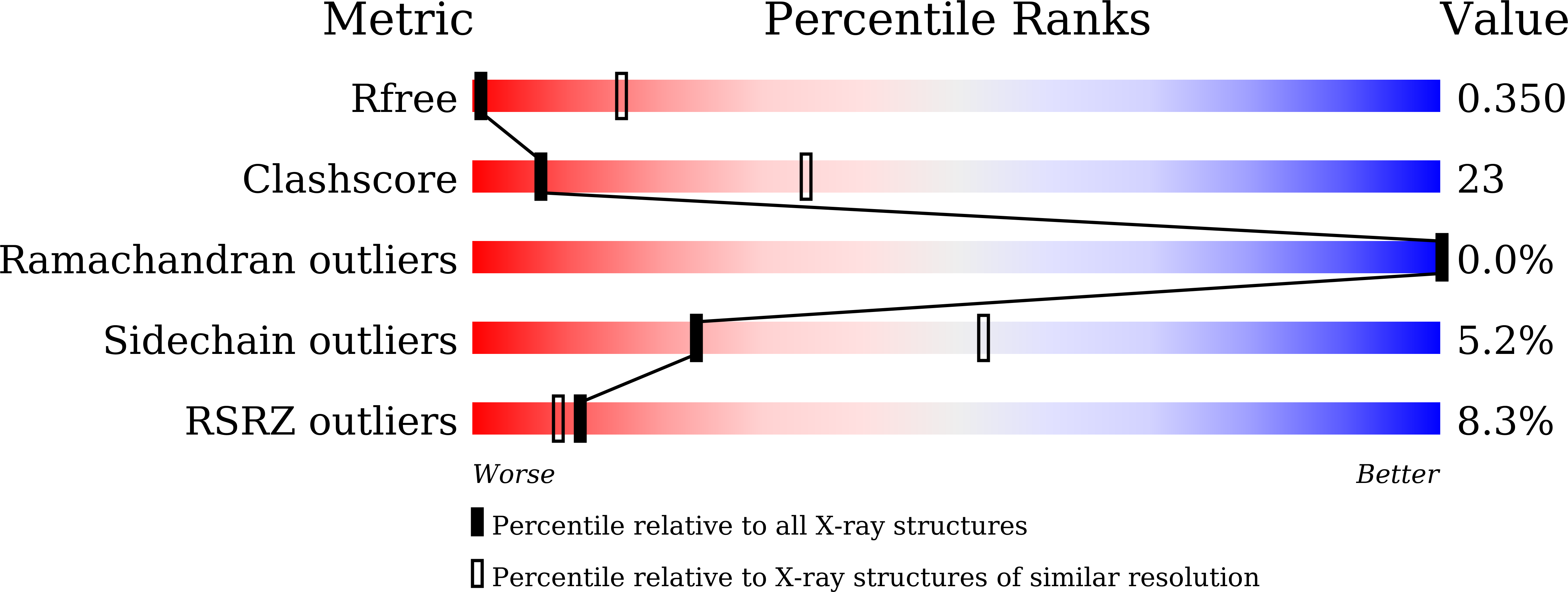
  • R-Value Free: 0.350 
  • R-Value Work: 0.281 
  • R-Value Observed: 0.285 

Starting Model: experimental
View more details

wwPDB Validation   3D Report Full Report


Ligand Structure Quality Assessment 


This is version 1.5 of the entry. See complete history


Literature

Crystal structure of a human plasma membrane phospholipid flippase.

Nakanishi, H.Irie, K.Segawa, K.Hasegawa, K.Fujiyoshi, Y.Nagata, S.Abe, K.

(2020) J Biol Chem 295: 10180-10194

  • DOI: https://doi.org/10.1074/jbc.RA120.014144
  • Primary Citation of Related Structures:  
    6LKN

  • PubMed Abstract: 

    ATP11C, a member of the P4-ATPase flippase, translocates phosphatidylserine from the outer to the inner plasma membrane leaflet, and maintains the asymmetric distribution of phosphatidylserine in the living cell. We present the crystal structures of a human plasma membrane flippase, ATP11C-CDC50A complex, in a stabilized E2P conformation. The structure revealed a deep longitudinal crevice along transmembrane helices continuing from the cell surface to the phospholipid occlusion site in the middle of the membrane. We observed that the extension of the crevice on the exoplasmic side is open, and the complex is therefore in an outward-open E2P state, similar to a recently reported cryo-EM structure of yeast flippase Drs2p-Cdc50p complex. We noted extra densities, most likely bound phosphatidylserines, in the crevice and in its extension to the extracellular side. One was close to the phosphatidylserine occlusion site as previously reported for the human ATP8A1-CDC50A complex, and the other in a cavity at the surface of the exoplasmic leaflet of the bilayer. Substitutions in either of the binding sites or along the path between them impaired specific ATPase and transport activities. These results provide evidence that the observed crevice is the conduit along that phosphatidylserine traverses from the outer leaflet to its occlusion site in the membrane and suggest that the exoplasmic cavity is important for phospholipid recognition. They also yield insights into how phosphatidylserine is incorporated from the outer leaflet of the plasma membrane into the transmembrane.


  • Organizational Affiliation

    Cellular and Structural Physiology Institute, Nagoya University, Nagoya, Japan.


Macromolecules
Find similar proteins by:  (by identity cutoff)  |  3D Structure
Entity ID: 1
MoleculeChains Sequence LengthOrganismDetailsImage
Phospholipid-transporting ATPase IGA,
C [auth E],
E [auth I],
G [auth M]
1,129Homo sapiensMutation(s): 1 
Gene Names: ATP11CATPIGATPIQ
EC: 7.6.2.1
Membrane Entity: Yes 
UniProt & NIH Common Fund Data Resources
Find proteins for Q8NB49 (Homo sapiens)
Explore Q8NB49 
Go to UniProtKB:  Q8NB49
PHAROS:  Q8NB49
GTEx:  ENSG00000101974 
Entity Groups  
Sequence Clusters30% Identity50% Identity70% Identity90% Identity95% Identity100% Identity
UniProt GroupQ8NB49
Sequence Annotations
Expand
  • Reference Sequence
Find similar proteins by:  (by identity cutoff)  |  3D Structure
Entity ID: 2
MoleculeChains Sequence LengthOrganismDetailsImage
Cell cycle control protein 50AB [auth C],
D [auth F],
F [auth J],
H [auth N]
361Homo sapiensMutation(s): 2 
Gene Names: TMEM30AC6orf67CDC50A
Membrane Entity: Yes 
UniProt & NIH Common Fund Data Resources
Find proteins for Q9NV96 (Homo sapiens)
Explore Q9NV96 
Go to UniProtKB:  Q9NV96
PHAROS:  Q9NV96
GTEx:  ENSG00000112697 
Entity Groups  
Sequence Clusters30% Identity50% Identity70% Identity90% Identity95% Identity100% Identity
UniProt GroupQ9NV96
Sequence Annotations
Expand
  • Reference Sequence
Small Molecules
Ligands 4 Unique
IDChains Name / Formula / InChI Key2D Diagram3D Interactions
P5S (Subject of Investigation/LOI)
Query on P5S

Download Ideal Coordinates CCD File 
GA [auth M]
I [auth A]
K [auth C]
Q [auth E]
R [auth E]
GA [auth M],
I [auth A],
K [auth C],
Q [auth E],
R [auth E],
Y [auth I],
Z [auth I]
O-[(R)-{[(2R)-2,3-bis(octadecanoyloxy)propyl]oxy}(hydroxy)phosphoryl]-L-serine
C42 H82 N O10 P
TZCPCKNHXULUIY-RGULYWFUSA-N
NAG
Query on NAG

Download Ideal Coordinates CCD File 
BA [auth J]
CA [auth J]
EA [auth J]
FA [auth J]
IA [auth N]
BA [auth J],
CA [auth J],
EA [auth J],
FA [auth J],
IA [auth N],
JA [auth N],
L [auth C],
LA [auth N],
M [auth C],
MA [auth N],
O [auth C],
P [auth C],
T [auth F],
U [auth F],
W [auth F],
X [auth F]
2-acetamido-2-deoxy-beta-D-glucopyranose
C8 H15 N O6
OVRNDRQMDRJTHS-FMDGEEDCSA-N
AH2
Query on AH2

Download Ideal Coordinates CCD File 
DA [auth J],
KA [auth N],
N [auth C],
V [auth F]
1-deoxy-alpha-D-mannopyranose
C6 H12 O5
MPCAJMNYNOGXPB-KVTDHHQDSA-N
MG
Query on MG

Download Ideal Coordinates CCD File 
AA [auth I],
HA [auth M],
J [auth A],
S [auth E]
MAGNESIUM ION
Mg
JLVVSXFLKOJNIY-UHFFFAOYSA-N
Modified Residues  1 Unique
IDChains TypeFormula2D DiagramParent
BFD
Query on BFD
A,
C [auth E],
E [auth I],
G [auth M]
L-PEPTIDE LINKINGC4 H6 Be F3 N O4ASP
Experimental Data & Validation

Experimental Data

  • Method: X-RAY DIFFRACTION
  • Resolution: 3.90 Å
  • R-Value Free: 0.350 
  • R-Value Work: 0.281 
  • R-Value Observed: 0.285 
  • Space Group: P 21 21 21
Unit Cell:
Length ( Å )Angle ( ˚ )
a = 100.46α = 90
b = 232.83β = 90
c = 492.89γ = 90
Software Package:
Software NamePurpose
PHENIXrefinement
PHENIXmodel building
XDSdata reduction
XDSdata scaling
PHASERphasing

Structure Validation

View Full Validation Report


Ligand Structure Quality Assessment 


Entry History & Funding Information

Deposition Data


Funding OrganizationLocationGrant Number
Japan Science and TechnologyJapanJPMJCR14M4
Japan Society for the Promotion of Science (JSPS)Japan17H03653

Revision History  (Full details and data files)

  • Version 1.0: 2020-06-10
    Type: Initial release
  • Version 1.1: 2020-06-17
    Changes: Database references
  • Version 1.2: 2020-07-15
    Changes: Structure summary
  • Version 1.3: 2020-07-29
    Type: Remediation
    Reason: Carbohydrate remediation
    Changes: Data collection, Derived calculations, Structure summary
  • Version 1.4: 2020-08-05
    Changes: Database references
  • Version 1.5: 2023-11-22
    Changes: Data collection, Database references, Refinement description, Structure summary