6LKC | pdb_00006lkc

Crystal structure of PfaD from Shewanella piezotolerans in complex with FMN


Experimental Data Snapshot

  • Method: X-RAY DIFFRACTION
  • Resolution: 2.00 Å
  • R-Value Free: 
    0.219 (Depositor), 0.225 (DCC) 
  • R-Value Work: 
    0.179 (Depositor), 0.192 (DCC) 
  • R-Value Observed: 
    0.181 (Depositor) 

Starting Model: experimental
View more details

wwPDB Validation 3D Report Full Report

Validation slider image for 6LKC

Ligand Structure Quality Assessment 


This is version 1.2 of the entry. See complete history

Literature

Structural Insights into the Trans -Acting Enoyl Reductase in the Biosynthesis of Long-Chain Polyunsaturated Fatty Acids in Shewanella piezotolerans .

Zhang, M.Zhang, H.Li, Q.Gao, Y.Guo, L.He, L.Zang, S.Guo, X.Huang, J.Li, L.

(2021) J Agric Food Chem 69: 2316-2324

  • DOI: https://doi.org/10.1021/acs.jafc.0c07386
  • Primary Citation Related Structures: 
    6LKC

  • PubMed Abstract: 

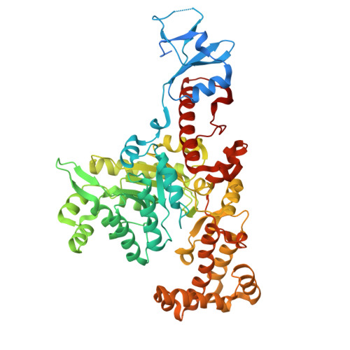
    Two long-chain polyunsaturated fatty acids (LC-PUFAs), eicosapentaenoic acid (EPA) and docosahexaenoic acid (DHA), play vital roles in human health. Similarly, two biosynthetic pathways, based on desaturase/elongase and polyketide synthase, have been implicated in the synthesis of microbial LC-PUFA. Up to now, only several microalgae, no bacteria, have been used in the commercial production of oils rich in DHA and/or EPA. Fully understanding the enzymatic mechanism in the biosynthesis of LC-PUFA would contribute significantly to produce EPA and/or DHA by the bacteria. In this study, we report a 1.998 Å-resolution crystal structure of trans -acting enoyl reductase (ER), SpPfaD, from Shewanella piezotolerans . The SpPfaD model consists of one homodimer in the asymmetric unit, and each subunit contains three domains. These include an N-terminal, a central domain forming a classic TIM barrel with a single FMN cofactor molecule bound atop the barrel, and a C-terminal domain with a lid above the TIM barrel. Furthermore, we docked oxidized nicotinamide adenine dinucleotide phosphate (NADP) and an inhibitor 2-(4-(2-((3-(5-(pyridin-2-ylthio)thiazol-2-yl)ureido)methyl)-1 H -imidazole-4-yl)phenoxy)acetic acid (TUI) molecule into the active site and analyzed the inhibition and catalytic mechanisms of the enoyl reductase SpPfaD. To the best of our knowledge, this is the first crystal structure of trans -ER in the biosynthesis of bacterial polyketides.


  • Organizational Affiliation
    • Engineering Research Center of Industrial Microbiology, Ministry of Education; Collaborative Innovation Center of Haixi Green Bio-Manufacturing Technology, Ministry of Education; National & Local Joint Engineering Research Center of Industrial Microbiology and Fermentation Technology, National Development and Reform Commission; College of Life Sciences, Fujian Normal University, Fuzhou, 350117, P. R. China.

Macromolecule Content 

  • Total Structure Weight: 130.11 kDa 
  • Atom Count: 8,766 
  • Modeled Residue Count: 1,063 
  • Deposited Residue Count: 1,174 
  • Unique protein chains: 1

Macromolecules

Find similar proteins by:|  3D Structure
Entity ID: 1
MoleculeChains  Sequence LengthOrganismDetailsImage
Polyunsaturated fatty acid synthase PfaD
A, B
587Shewanella piezotolerans WP3Mutation(s): 0 
Gene Names: swp_3554
EC: 1.3.1.9
UniProt
Find proteins for B8CQB6 (Shewanella piezotolerans (strain WP3 / JCM 13877))
Explore B8CQB6 
Go to UniProtKB:  B8CQB6
Entity Groups
Sequence Clusters30% Identity50% Identity70% Identity90% Identity95% Identity100% Identity
UniProt GroupB8CQB6
Sequence Annotations
Expand
Reference Sequence

Small Molecules

Ligands 3 Unique
IDChains Name / Formula / InChI Key2D Diagram3D Interactions
FMN

Query on FMN


Download:Ideal Coordinates CCD File
C [auth A],
F [auth B]
FLAVIN MONONUCLEOTIDE
C17 H21 N4 O9 P
FVTCRASFADXXNN-SCRDCRAPSA-N
GOL

Query on GOL


Download:Ideal Coordinates CCD File
E [auth A],
H [auth B],
I [auth B]
GLYCEROL
C3 H8 O3
PEDCQBHIVMGVHV-UHFFFAOYSA-N
CA

Query on CA


Download:Ideal Coordinates CCD File
D [auth A],
G [auth B]
CALCIUM ION
Ca
BHPQYMZQTOCNFJ-UHFFFAOYSA-N

Experimental Data & Validation

Experimental Data

  • Method: X-RAY DIFFRACTION
  • Resolution: 2.00 Å
  • R-Value Free:  0.219 (Depositor), 0.225 (DCC) 
  • R-Value Work:  0.179 (Depositor), 0.192 (DCC) 
  • R-Value Observed: 0.181 (Depositor) 
Space Group: P 1 21 1
Unit Cell:
Length ( Å )Angle ( ˚ )
a = 70.467α = 90
b = 62.568β = 105.61
c = 127.507γ = 90
Software Package:
Software NamePurpose
PHENIXrefinement
PDB_EXTRACTdata extraction
XDSdata reduction
Aimlessdata scaling
PHASERphasing

Structure Validation

View Full Validation Report


Ligand Structure Quality Assessment 


Entry History 

Deposition Data

Revision History  (Full details and data files)

  • Version 1.0: 2020-12-23
    Type: Initial release
  • Version 1.1: 2021-04-07
    Changes: Database references
  • Version 1.2: 2023-11-22
    Changes: Data collection, Database references, Refinement description