


Crystal structure of PCB4scFv(hN56D)
Nakamura, T., Morioka, H.To be published.
Experimental Data Snapshot
Starting Model: experimental
View more details
wwPDB Validation 3D Report Full Report

Entity ID: 1 | |||||
|---|---|---|---|---|---|
| Molecule | Chains | Sequence Length | Organism | Details | Image |
| PCB4scFv(hN56D) | 253 | Mus musculus | Mutation(s): 0 | 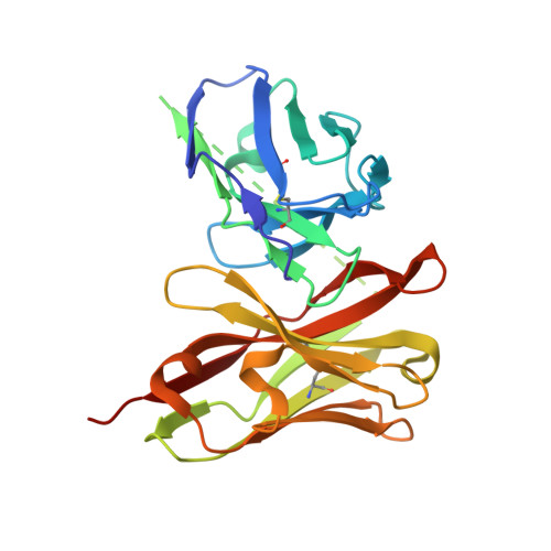 | |
| Length ( Å ) | Angle ( ˚ ) |
|---|---|
| a = 92.42 | α = 90 |
| b = 92.42 | β = 90 |
| c = 53.83 | γ = 90 |
| Software Name | Purpose |
|---|---|
| PHENIX | refinement |
| XDS | data reduction |
| XDS | data scaling |
| MOLREP | phasing |
| Funding Organization | Location | Grant Number |
|---|---|---|
| Ministry of Education, Culture, Sports, Science and Technology (Japan) | Japan | -- |


