


Crystal Structure of Pluripotency Reprogramming Factor Sox17 mutant (Sox17EK) HMG Domain bound to DNA
Balasubramanian, M., Kolatkar, P.R.To be published.
Experimental Data Snapshot
Starting Model: experimental
View more details
Entity ID: 3 | |||||
|---|---|---|---|---|---|
| Molecule | Chains | Sequence Length | Organism | Details | Image |
| Transcription factor SOX-17 | C [auth D], F | 83 | Mus musculus | Mutation(s): 1 Gene Names: Sox17, Sox-17 | 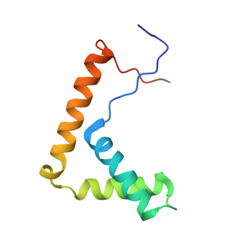 |
UniProt | |||||
Entity Groups | |||||
| Sequence Clusters | 30% Identity50% Identity70% Identity90% Identity95% Identity100% Identity | ||||
| UniProt Group | Q61473 | ||||
Sequence AnnotationsExpand | |||||
Reference Sequence | |||||
Entity ID: 1 | ||||
| Molecule | Chains | Length | Organism | Image |
|---|---|---|---|---|
| DNA (5'-D(P*GP*GP*TP*CP*TP*CP*TP*AP*TP*TP*GP*TP*CP*CP*TP*G)-3') | A, D [auth C] | 16 | Homo sapiens |  |
Sequence AnnotationsExpand | ||||
Reference Sequence | ||||
Entity ID: 2 | ||||
| Molecule | Chains | Length | Organism | Image |
|---|---|---|---|---|
| DNA (5'-D(*CP*CP*AP*GP*GP*AP*CP*AP*AP*TP*AP*GP*AP*GP*AP*C)-3') | 16 | Homo sapiens | 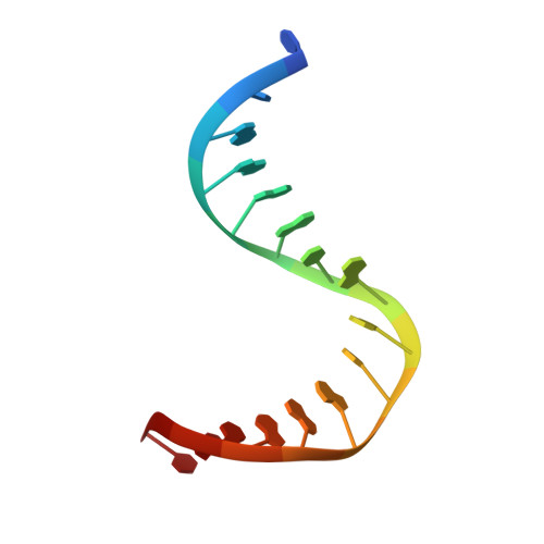 | |
Sequence AnnotationsExpand | ||||
Reference Sequence | ||||
| Ligands 1 Unique | |||||
|---|---|---|---|---|---|
| ID | Chains | Name / Formula / InChI Key | 2D Diagram | 3D Interactions | |
| SO4 (Subject of Investigation/LOI) Download:Ideal Coordinates CCD File | G [auth D] | SULFATE ION O4 S QAOWNCQODCNURD-UHFFFAOYSA-L |  | ||
| Length ( Å ) | Angle ( ˚ ) |
|---|---|
| a = 71.04 | α = 90 |
| b = 73.205 | β = 90 |
| c = 81.952 | γ = 90 |
| Software Name | Purpose |
|---|---|
| PHENIX | refinement |
| PROTEUM | data reduction |
| SADABS | data scaling |
| PHENIX | phasing |
| PROTEUM | data collection |
| Funding Organization | Location | Grant Number |
|---|---|---|
| Qatar Foundation | Qatar | IGP1 2014-004 |


