


Structural Analysis of Saccharomyces cerevisiae Dihydroorotase Reveals Molecular Insights into the Tetramerization Mechanism
Guan, H.H., Huang, Y.H., Lin, E.S., Chen, C.J., Huang, C.Y.(2021) Molecules
Experimental Data Snapshot
(2021) Molecules
Entity ID: 1 | |||||
|---|---|---|---|---|---|
| Molecule | Chains | Sequence Length | Organism | Details | Image |
| Dihydroorotase | 372 | Saccharomyces cerevisiae S288C | Mutation(s): 0 Gene Names: URA4, YLR420W, L9931.1 EC: 3.5.2.3 | 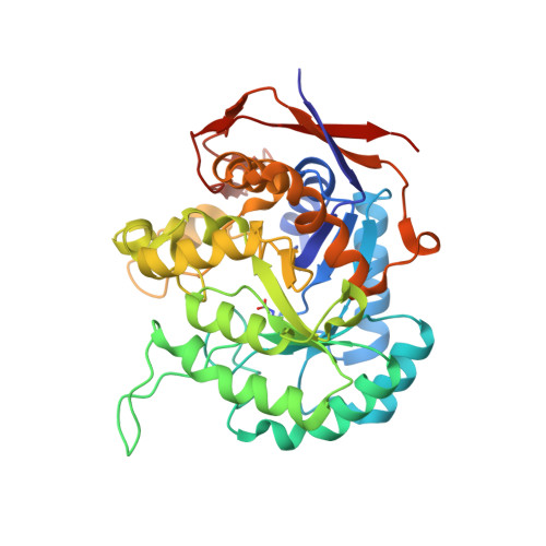 | |
UniProt | |||||
Entity Groups | |||||
| Sequence Clusters | 30% Identity50% Identity70% Identity90% Identity95% Identity100% Identity | ||||
| UniProt Group | P20051 | ||||
Sequence AnnotationsExpand | |||||
Reference Sequence | |||||
| Ligands 2 Unique | |||||
|---|---|---|---|---|---|
| ID | Chains | Name / Formula / InChI Key | 2D Diagram | 3D Interactions | |
| LMR (Subject of Investigation/LOI) Download:Ideal Coordinates CCD File | G [auth A], J [auth B], M [auth C], P [auth D] | (2S)-2-hydroxybutanedioic acid C4 H6 O5 BJEPYKJPYRNKOW-REOHCLBHSA-N |  | ||
| ZN Download:Ideal Coordinates CCD File | E [auth A] F [auth A] H [auth B] I [auth B] K [auth C] | ZINC ION Zn PTFCDOFLOPIGGS-UHFFFAOYSA-N |  | ||
| Modified Residues 1 Unique | |||||
|---|---|---|---|---|---|
| ID | Chains | Type | Formula | 2D Diagram | Parent |
| KCX Query on KCX | A, B, C, D | L-PEPTIDE LINKING | C7 H14 N2 O4 |  | LYS |
| Length ( Å ) | Angle ( ˚ ) |
|---|---|
| a = 85.568 | α = 90 |
| b = 88.544 | β = 95.73 |
| c = 103.079 | γ = 90 |
| Software Name | Purpose |
|---|---|
| PHENIX | refinement |
| PDB_EXTRACT | data extraction |
| Epinorm | data reduction |
| HKL-2000 | data scaling |
| AutoSol | phasing |
| Funding Organization | Location | Grant Number |
|---|---|---|
| Ministry of Science and Technology (Taiwan) | Taiwan | -- |


