6KZI

Crystal structure of Ser/Thr kinase Pim1 in complex with thioridazine derivatives


Experimental Data Snapshot

  • Method: X-RAY DIFFRACTION
  • Resolution: 2.80 Å
  • R-Value Free: 
    0.233 (Depositor), 0.230 (DCC) 
  • R-Value Work: 
    0.170 (Depositor), 0.180 (DCC) 
  • R-Value Observed: 
    0.173 (Depositor) 

wwPDB Validation   3D Report Full Report


Ligand Structure Quality Assessment 

Created with Raphaël 2.3.0Worse 01 BetterLigand structure goodness of fit to experimental dataBest fitted RTXClick on this verticalbar to view details

This is version 1.2 of the entry. See complete history


Literature

Structure-Based Optimization of 10-DEBC Derivatives as Potent and Selective Pim-1 Kinase Inhibitors.

Li, G.Zhang, W.Xie, Y.Li, Y.Cao, R.Zheng, G.Huang, N.Zhou, Y.

(2020) J Chem Inf Model 60: 3287-3294

  • DOI: https://doi.org/10.1021/acs.jcim.0c00245
  • Primary Citation of Related Structures:  
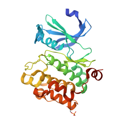
    6KZI, 6L11, 6L12, 6L13, 6L14, 6L15, 6L16, 6L17

  • PubMed Abstract: 

    Pim-1 kinase has been widely regarded as an attractive target for anticancer drugs. Here, we reported our continued efforts in structure-based optimization of compound 10-DEBC, a previously identified micromolar Pim-1 inhibitor. Guided by the Site Identification by Ligand Competitive Saturation (SILCS) method, we quickly obtained a series of 10-DEBC derivatives with significantly improved activity and selectivity. In particular, compound 26 exhibited an IC 50 value of 0.9 nM, as well as 220- and 8-fold selectivity over Pim-2 and Pim-3 kinases, respectively.


  • Organizational Affiliation

    State Key Laboratory of Chemical Resources Engineering, Beijing University of Chemical Technology, Beijing 100029, China.


Macromolecules
Find similar proteins by:  (by identity cutoff)  |  3D Structure
Entity ID: 1
MoleculeChains Sequence LengthOrganismDetailsImage
Serine/threonine-protein kinase pim-1289Homo sapiensMutation(s): 0 
Gene Names: PIM1
EC: 2.7.11.1
UniProt & NIH Common Fund Data Resources
Find proteins for P11309 (Homo sapiens)
Explore P11309 
Go to UniProtKB:  P11309
PHAROS:  P11309
GTEx:  ENSG00000137193 
Entity Groups  
Sequence Clusters30% Identity50% Identity70% Identity90% Identity95% Identity100% Identity
UniProt GroupP11309
Sequence Annotations
Expand
  • Reference Sequence
Small Molecules
Ligands 1 Unique
IDChains Name / Formula / InChI Key2D Diagram3D Interactions
RTX (Subject of Investigation/LOI)
Query on RTX

Download Ideal Coordinates CCD File 
B [auth A]4-(2-chloro-10H-phenoxazin-10-yl)-N,N-diethylbutan-1-amine
C20 H25 Cl N2 O
GYBXAGDWMCJZJK-UHFFFAOYSA-N
Modified Residues  1 Unique
IDChains TypeFormula2D DiagramParent
SEP
Query on SEP
A
L-PEPTIDE LINKINGC3 H8 N O6 PSER
Experimental Data & Validation

Experimental Data

  • Method: X-RAY DIFFRACTION
  • Resolution: 2.80 Å
  • R-Value Free:  0.233 (Depositor), 0.230 (DCC) 
  • R-Value Work:  0.170 (Depositor), 0.180 (DCC) 
  • R-Value Observed: 0.173 (Depositor) 
Space Group: P 65
Unit Cell:
Length ( Å )Angle ( ˚ )
a = 98.925α = 90
b = 98.925β = 90
c = 80.83γ = 120
Software Package:
Software NamePurpose
REFMACrefinement
PDB_EXTRACTdata extraction
HKL-2000data reduction
SCALEPACKdata scaling
PHASESphasing

Structure Validation

View Full Validation Report


Ligand Structure Quality Assessment 

Created with Raphaël 2.3.0Worse 01 BetterLigand structure goodness of fit to experimental dataBest fitted RTXClick on this verticalbar to view details

Entry History & Funding Information

Deposition Data


Funding OrganizationLocationGrant Number
Chinese Academy of SciencesChina2011CB812402

Revision History  (Full details and data files)

  • Version 1.0: 2020-03-04
    Type: Initial release
  • Version 1.1: 2021-03-17
    Changes: Database references
  • Version 1.2: 2024-11-06
    Changes: Data collection, Database references, Structure summary