6KV9 | pdb_00006kv9

MoeE5 in complex with UDP-glucuronic acid and NAD


Experimental Data Snapshot

  • Method: X-RAY DIFFRACTION
  • Resolution: 1.48 Å
  • R-Value Free: 
    0.186 (Depositor), 0.184 (DCC) 
  • R-Value Work: 
    0.159 (Depositor), 0.159 (DCC) 
  • R-Value Observed: 
    0.160 (Depositor) 

Starting Model: experimental
View more details

wwPDB Validation 3D Report Full Report

Validation slider image for 6KV9

Ligand Structure Quality Assessment 


This is version 1.2 of the entry. See complete history

Literature

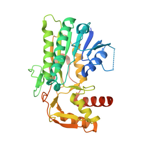
Structure of an antibiotic-synthesizing UDP-glucuronate 4-epimerase MoeE5 in complex with substrate.

Sun, H.Ko, T.P.Liu, W.Liu, W.Zheng, Y.Chen, C.C.Guo, R.T.

(2020) Biochem Biophys Res Commun 521: 31-36

  • DOI: https://doi.org/10.1016/j.bbrc.2019.10.035
  • Primary Citation Related Structures: 
    6KV9, 6KVC

  • PubMed Abstract: 

    The epimerase MoeE5 from Streptomyces viridosporus converts UDP-glucuronic acid (UDP-GlcA) to UDP-galacturonic acid (UDP-GalA) to provide the first sugar in synthesizing moenomycin, a potent inhibitor against bacterial peptidoglycan glycosyltransferases. The enzyme belongs to the UDP-hexose 4-epimerase family, and uses NAD + as its cofactor. Here we present the complex crystal structures of MoeE5/NAD + /UDP-GlcA and MoeE5/NAD + /UDP-glucose, determined at 1.48 Å and 1.66 Å resolution. The cofactor NAD + is bound to the N-terminal Rossmann-fold domain and the substrate is bound to the smaller C-terminal domain. In both crystals the C4 atom of the sugar moiety of the substrate is in close proximity to the C4 atom of the nicotinamide of NAD + , and the O4 atom of the sugar is also hydrogen bonded to the side chain of Tyr154, suggesting a productive binding mode. As the first complex structure of this protein family with a bound UDP-GlcA in the active site, it shows an extensive hydrogen-bond network between the enzyme and the substrate. We further built a model with the product UDP-GalA, and found that the unique Arg192 of MoeE5 might play an important role in the catalytic pathway. Consequently, MoeE5 is likely a specific epimerase for UDP-GlcA to UDP-GalA conversion, rather than a promiscuous enzyme as some other family members.


  • Organizational Affiliation
    • University of Chinese Academy of Sciences, Beijing, 100049, China; Tianjin Institute of Industrial Biotechnology, Chinese Academy of Sciences, Tianjin, 300308, China.

Macromolecule Content 

  • Total Structure Weight: 37.79 kDa 
  • Atom Count: 2,786 
  • Modeled Residue Count: 299 
  • Deposited Residue Count: 346 
  • Unique protein chains: 1

Macromolecules

Find similar proteins by:|  3D Structure
Entity ID: 1
MoleculeChains  Sequence LengthOrganismDetailsImage
MoeE5346Streptomyces viridosporus ATCC 14672Mutation(s): 0 
Gene Names: moeE5
UniProt
Find proteins for A0A003 (Streptomyces viridosporus)
Explore A0A003 
Go to UniProtKB:  A0A003
Entity Groups
Sequence Clusters30% Identity50% Identity70% Identity90% Identity95% Identity100% Identity
UniProt GroupA0A003
Sequence Annotations
Expand
Reference Sequence

Small Molecules

Experimental Data & Validation

Experimental Data

  • Method: X-RAY DIFFRACTION
  • Resolution: 1.48 Å
  • R-Value Free:  0.186 (Depositor), 0.184 (DCC) 
  • R-Value Work:  0.159 (Depositor), 0.159 (DCC) 
  • R-Value Observed: 0.160 (Depositor) 
Space Group: P 41 21 2
Unit Cell:
Length ( Å )Angle ( ˚ )
a = 59.159α = 90
b = 59.159β = 90
c = 200.387γ = 90
Software Package:
Software NamePurpose
PHENIXrefinement
HKL-2000data reduction
HKL-2000data scaling
PHASERphasing

Structure Validation

View Full Validation Report


Ligand Structure Quality Assessment 


Entry History 

Deposition Data

Revision History  (Full details and data files)

  • Version 1.0: 2019-11-13
    Type: Initial release
  • Version 1.1: 2020-01-08
    Changes: Database references
  • Version 1.2: 2023-11-22
    Changes: Data collection, Database references, Refinement description