Funding Organization(s): Japan Agency for Medical Research and Development (AMED), Ministry of Education, Culture, Sports, Science and Technology (Japan)
Primary Citation Related Structures: 6KRZ, 6KS0, 6KS1
PubMed Abstract:
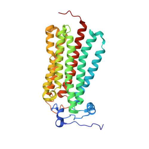
The human adiponectin receptors, AdipoR1 and AdipoR2, are key anti-diabetic molecules. We previously reported the crystal structures of human AdipoR1 and AdipoR2, revealing that their seven transmembrane helices form an internal closed cavity (the closed form). In this study, we determined the crystal structure of the D208A variant AdipoR1, which is fully active with respect to the major downstream signaling. Among the three molecules in the asymmetric unit, two assume the closed form, and the other adopts the open form with large openings in the internal cavity. Between the closed- and open-form structures, helices IV and V are tilted with their intracellular ends shifted by about 4 and 11 Å, respectively. Furthermore, we reanalyzed our previous wild-type AdipoR1 diffraction data, and determined a 44:56 mixture of the closed and open forms, respectively. Thus, we have clarified the closed-open interconversion of AdipoR1, which may be relevant to its functional mechanism(s).
RIKEN Cluster for Science, Technology and Innovation Hub, 1-7-22 Suehiro-cho, Tsurumi-ku, Yokohama, 230-0045, Japan.
Division of Structural and Synthetic Biology, RIKEN Center for Life Science Technologies, 1-7-22 Suehiro-cho, Tsurumi-ku, Yokohama, 230-0045, Japan.
Department of Diabetes and Metabolic Diseases, Graduate School of Medicine, The University of Tokyo, 7-3-1 Hongo, Bunkyo-ku, Tokyo, 113-0033, Japan.
Laboratory for Advanced Research on Pathophysiology of Metabolic Diseases, The University of Tokyo, 7-3-1 Hongo, Bunkyo-ku, Tokyo, 113-0033, Japan.
Department of Integrated Molecular Sciences on Metabolic Diseases, 22nd Century Medical and Research Center, The University of Tokyo, 7-3-1 Hongo, Bunkyo-ku, Tokyo, 113-0033, Japan.
Laboratory of Molecular and Cellular Biochemistry, Graduate School of Pharmaceutical Sciences, Tohoku University, Miyagi, 980-8578, Japan.
AMED-CREST, Japan Agency for Medical Research and Development, Otemachi, Chiyoda-ku, Tokyo, 100-0004, Japan.
Laboratory for Protein Functional and Structural Biology, RIKEN Center for Biosystems Dynamics Research, 1-7-22 Suehiro-cho, Tsurumi-ku, Yokohama, 230-0045, Japan.
RIKEN SPring-8 Center, Kouto, Sayo, Hyogo, 679-5148, Japan.
Department of Diabetes and Metabolic Diseases, Graduate School of Medicine, The University of Tokyo, 7-3-1 Hongo, Bunkyo-ku, Tokyo, 113-0033, Japan. tyamau-tky@umin.net.
Laboratory for Advanced Research on Pathophysiology of Metabolic Diseases, The University of Tokyo, 7-3-1 Hongo, Bunkyo-ku, Tokyo, 113-0033, Japan. tyamau-tky@umin.net.
Department of Integrated Molecular Sciences on Metabolic Diseases, 22nd Century Medical and Research Center, The University of Tokyo, 7-3-1 Hongo, Bunkyo-ku, Tokyo, 113-0033, Japan. tyamau-tky@umin.net.
AMED-CREST, Japan Agency for Medical Research and Development, Otemachi, Chiyoda-ku, Tokyo, 100-0004, Japan. tyamau-tky@umin.net.
Department of Diabetes and Metabolic Diseases, Graduate School of Medicine, The University of Tokyo, 7-3-1 Hongo, Bunkyo-ku, Tokyo, 113-0033, Japan. kadowaki-3im@h.u-tokyo.ac.jp.
Laboratory for Advanced Research on Pathophysiology of Metabolic Diseases, The University of Tokyo, 7-3-1 Hongo, Bunkyo-ku, Tokyo, 113-0033, Japan. kadowaki-3im@h.u-tokyo.ac.jp.
Department of Integrated Molecular Sciences on Metabolic Diseases, 22nd Century Medical and Research Center, The University of Tokyo, 7-3-1 Hongo, Bunkyo-ku, Tokyo, 113-0033, Japan. kadowaki-3im@h.u-tokyo.ac.jp.
Department of Metabolism and Nutrition, Mizonokuchi Hospital, Faculty of Medicine, Teikyo University, 5-1-1 Futago, Takatsu-ku, Kawasaki, Kanagawa, 213-8507, Japan. kadowaki-3im@h.u-tokyo.ac.jp.