6KNZ | pdb_00006knz

Crystal structure of T2R-TTL-KXO1 complex


Experimental Data Snapshot

  • Method: X-RAY DIFFRACTION
  • Resolution: 2.48 Å
  • R-Value Free: 
    0.261 (Depositor), 0.262 (DCC) 
  • R-Value Work: 
    0.220 (Depositor), 0.221 (DCC) 
  • R-Value Observed: 
    0.221 (Depositor) 

Starting Model: experimental
View more details

wwPDB Validation 3D Report Full Report

Validation slider image for 6KNZ

Ligand Structure Quality Assessment 


This is version 1.3 of the entry. See complete history

Literature

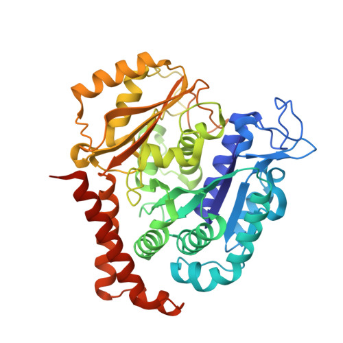
Reversible binding of the anticancer drug KXO1 (tirbanibulin) to the colchicine-binding site of beta-tubulin explains KXO1's low clinical toxicity.

Niu, L.Yang, J.Yan, W.Yu, Y.Zheng, Y.Ye, H.Chen, Q.Chen, L.

(2019) J Biological Chem 294: 18099-18108

  • DOI: https://doi.org/10.1074/jbc.RA119.010732
  • Primary Citation Related Structures: 
    6KNZ

  • PubMed Abstract: 

    KXO1 (tirbanibulin or KX2-391) is as a non-ATP-competitive inhibitor of SRC proto-oncogene nonreceptor tyrosine kinase (SRC) and is being clinically investigated for the management of various cancers and actinic keratosis. Recently, KXO1 has also been shown to strongly inhibit tubulin. Interestingly, unlike conventional tubulin-targeting drugs, KXO1 has exhibited low toxicity in preclinical and clinical studies, but the reason for this remains elusive, as are the KXO1-binding site and other details of the interaction of KXO1 with tubulin. Here, cell-based experiments revealed that KXO1 induces tubulin depolymerization and G 2 /M phase cell cycle arrest at low nanomolar concentrations, similar to colchicine, used as a positive control. Results from biochemical experiments, including an N , N -ethylenebis(iodoacetamide) competition assay, disclosed that KXO1 binds to the colchicine-binding site on β-tubulin, further confirmed by the crystal structure of the tubulin-KXO1 complex at 2.5-Å resolution. A high-quality electron density map of the crystallographic data enabled us to unambiguously determine the position and orientation of KXO1 in the colchicine-binding site, revealing the detailed interactions between KXO1 and tubulin. We also found that KXO1 binds reversibly to purified tubulin, induces a totally reversible cellular effect (G 2 /M cell cycle arrest), and possesses no cellular toxicity 5 days after drug washout, explaining KXO1's low toxicity. In summary, we show that KXO1 binds to the colchicine-binding site of tubulin and resolved the crystal structure of the tubulin-KXO1 complex. Importantly, KXO1's reversible binding to tubulin explains its clinically low toxicity, an insight that could guide further clinical applications of KXO1.


  • Organizational Affiliation
    • State Key Laboratory of Biotherapy and Cancer Center, West China Hospital of Sichuan University and Collaborative Innovation Center of Biotherapy and Cancer, Chengdu 610041, China.

Macromolecule Content 

  • Total Structure Weight: 265.44 kDa 
  • Atom Count: 17,969 
  • Modeled Residue Count: 2,200 
  • Deposited Residue Count: 2,317 
  • Unique protein chains: 4

Macromolecules

Find similar proteins by:|  3D Structure
Entity ID: 1
MoleculeChains  Sequence LengthOrganismDetailsImage
Tubulin alpha-1B chain
A, C
450Bos taurusMutation(s): 0 
EC: 3.6.5
UniProt
Find proteins for P81947 (Bos taurus)
Explore P81947 
Go to UniProtKB:  P81947
Entity Groups
Sequence Clusters30% Identity50% Identity70% Identity90% Identity95% Identity100% Identity
UniProt GroupP81947
Sequence Annotations
Expand
Reference Sequence
Find similar proteins by:|  3D Structure
Entity ID: 2
MoleculeChains  Sequence LengthOrganismDetailsImage
Tubulin beta-2B chain
B, D
445Bos taurusMutation(s): 0 
UniProt
Find proteins for Q6B856 (Bos taurus)
Explore Q6B856 
Go to UniProtKB:  Q6B856
Entity Groups
Sequence Clusters30% Identity50% Identity70% Identity90% Identity95% Identity100% Identity
UniProt GroupQ6B856
Sequence Annotations
Expand
Reference Sequence
Find similar proteins by:|  3D Structure
Entity ID: 3
MoleculeChains  Sequence LengthOrganismDetailsImage
Stathmin-4143Rattus norvegicusMutation(s): 0 
Gene Names: Stmn4
UniProt
Find proteins for P63043 (Rattus norvegicus)
Explore P63043 
Go to UniProtKB:  P63043
Entity Groups
Sequence Clusters30% Identity50% Identity70% Identity90% Identity95% Identity100% Identity
UniProt GroupP63043
Sequence Annotations
Expand
Reference Sequence
Find similar proteins by:|  3D Structure
Entity ID: 4
MoleculeChains  Sequence LengthOrganismDetailsImage
Tubulin tyrosine ligase384Gallus gallusMutation(s): 0 
Gene Names: TTL

Small Molecules

Ligands 7 Unique
IDChains Name / Formula / InChI Key2D Diagram3D Interactions
GTP

Query on GTP


Download:Ideal Coordinates CCD File
G [auth A],
Q [auth C],
V [auth D]
GUANOSINE-5'-TRIPHOSPHATE
C10 H16 N5 O14 P3
XKMLYUALXHKNFT-UUOKFMHZSA-N
ACP

Query on ACP


Download:Ideal Coordinates CCD File
Z [auth F]PHOSPHOMETHYLPHOSPHONIC ACID ADENYLATE ESTER
C11 H18 N5 O12 P3
UFZTZBNSLXELAL-IOSLPCCCSA-N
GDP

Query on GDP


Download:Ideal Coordinates CCD File
J [auth B]GUANOSINE-5'-DIPHOSPHATE
C10 H15 N5 O11 P2
QGWNDRXFNXRZMB-UUOKFMHZSA-N
DN0
(Subject of Investigation/LOI)

Query on DN0


Download:Ideal Coordinates CCD File
O [auth B],
W [auth D]
2-[5-[4-(2-morpholin-4-ylethoxy)phenyl]pyridin-2-yl]-~{N}-(phenylmethyl)ethanamide
C26 H29 N3 O3
HUNGUWOZPQBXGX-UHFFFAOYSA-N
MES

Query on MES


Download:Ideal Coordinates CCD File
L [auth B],
P [auth B]
2-(N-MORPHOLINO)-ETHANESULFONIC ACID
C6 H13 N O4 S
SXGZJKUKBWWHRA-UHFFFAOYSA-N
CA

Query on CA


Download:Ideal Coordinates CCD File
I [auth A]
M [auth B]
N [auth B]
S [auth C]
U [auth D]
I [auth A],
M [auth B],
N [auth B],
S [auth C],
U [auth D],
X [auth E]
CALCIUM ION
Ca
BHPQYMZQTOCNFJ-UHFFFAOYSA-N
MG

Query on MG


Download:Ideal Coordinates CCD File
H [auth A],
K [auth B],
R [auth C],
T [auth D],
Y [auth F]
MAGNESIUM ION
Mg
JLVVSXFLKOJNIY-UHFFFAOYSA-N

Experimental Data & Validation

Experimental Data

  • Method: X-RAY DIFFRACTION
  • Resolution: 2.48 Å
  • R-Value Free:  0.261 (Depositor), 0.262 (DCC) 
  • R-Value Work:  0.220 (Depositor), 0.221 (DCC) 
  • R-Value Observed: 0.221 (Depositor) 
Space Group: P 21 21 21
Unit Cell:
Length ( Å )Angle ( ˚ )
a = 104.928α = 90
b = 157.059β = 90
c = 182.21γ = 90
Software Package:
Software NamePurpose
HKL-2000data scaling
PHENIXrefinement
PDB_EXTRACTdata extraction
HKL-3000data reduction
HKL-3000phasing

Structure Validation

View Full Validation Report


Ligand Structure Quality Assessment 


Entry History 

Deposition Data

Revision History  (Full details and data files)

  • Version 1.0: 2019-10-23
    Type: Initial release
  • Version 1.1: 2019-10-30
    Changes: Data collection, Database references
  • Version 1.2: 2019-12-18
    Changes: Database references
  • Version 1.3: 2023-11-22
    Changes: Data collection, Database references, Derived calculations, Refinement description