6KHX

Crystal structure of Prx from Akkermansia muciniphila


Experimental Data Snapshot

  • Method: X-RAY DIFFRACTION
  • Resolution: 2.58 Å
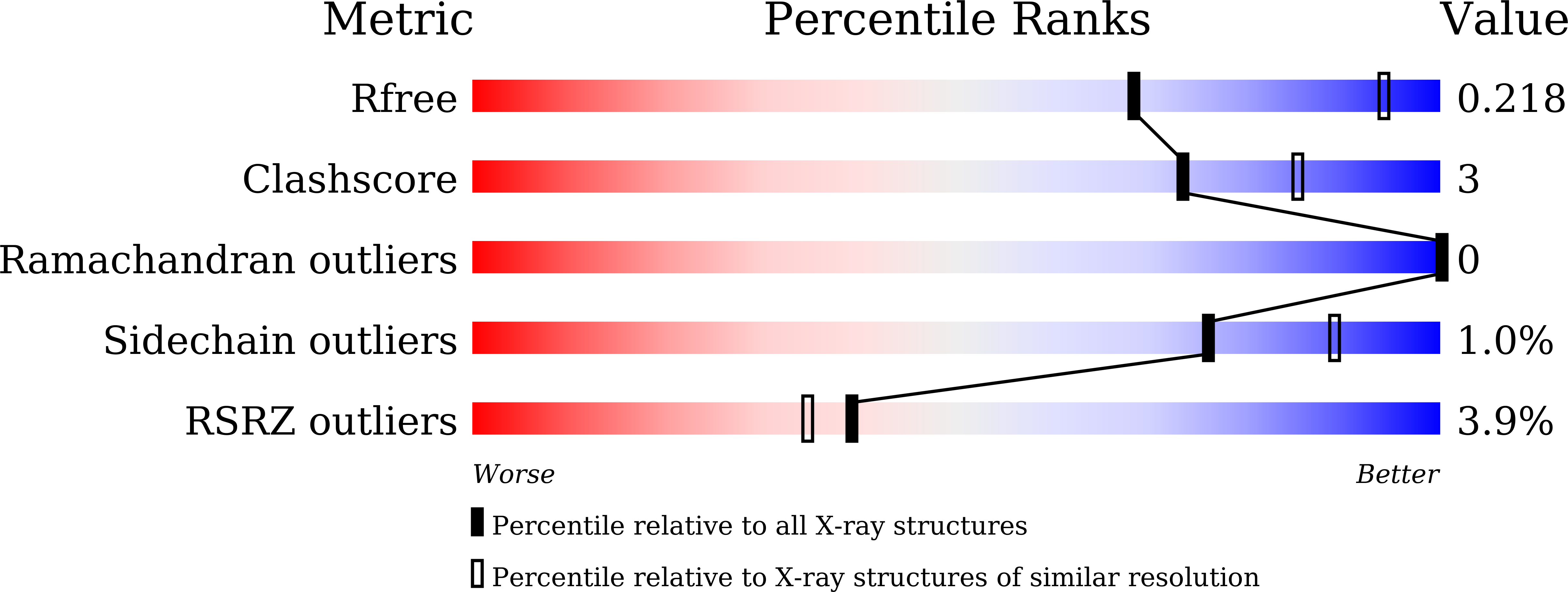
  • R-Value Free: 0.218 
  • R-Value Work: 0.177 
  • R-Value Observed: 0.179 

Starting Model: experimental
View more details

wwPDB Validation   3D Report Full Report


This is version 1.2 of the entry. See complete history


Literature

Crystal structure of Akkermansia muciniphila peroxiredoxin reveals a novel regulatory mechanism of typical 2-Cys Prxs by a distinct loop.

Li, M.Wang, J.Xu, W.Wang, Y.Zhang, M.Wang, M.

(2020) FEBS Lett 594: 1550-1563

  • DOI: https://doi.org/10.1002/1873-3468.13753
  • Primary Citation of Related Structures:  
    6KHX

  • PubMed Abstract: 

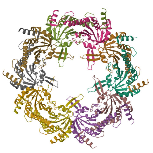
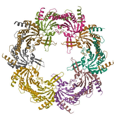
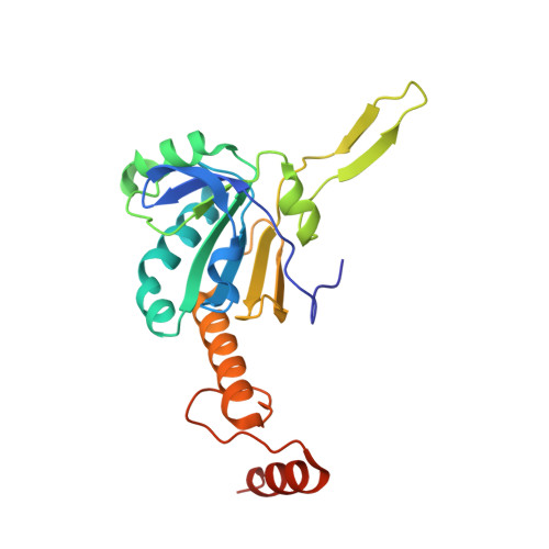
    Peroxiredoxins (Prxs) are thiol-specific antioxidant proteins commonly found in organisms that protect cells from the damage of reactive oxygen species produced by metabolism and that participate in cell signaling. The Prx from the bacterium Akkermansia muciniphila (AmPrx) is a typical 2-Cys Prx characterized by two conserved cysteines: Cys49 and Cys183. Here, we verified the peroxidase activity of AmPrx and determined its crystal structure in reduced form, which is a doughnut-shaped decamer composed of five dimers. Particularly, a distinct loop between the α4 helix and β6 strand is involved in the decameric interaction. Deletion of this loop destroys the decameric structure and significantly decreases the peroxidase activity of AmPrx. Our findings reveal a novel regulatory mechanism of typical 2-Cys Prx, in which the α4-β6 loop affects the assembly of Prx and, therefore, regulates its peroxidase activity.


  • Organizational Affiliation

    School of Life Sciences, Anhui University, Hefei, China.


Macromolecules
Find similar proteins by:  (by identity cutoff)  |  3D Structure
Entity ID: 1
MoleculeChains Sequence LengthOrganismDetailsImage
Peroxiredoxin
A, B, C, D, E
A, B, C, D, E, F, G, H, I, J
215Akkermansia muciniphilaMutation(s): 0 
Gene Names: CXU17_12910CXU19_10505
Entity Groups  
Sequence Clusters30% Identity50% Identity70% Identity90% Identity95% Identity100% Identity
Sequence Annotations
Expand
  • Reference Sequence
Experimental Data & Validation

Experimental Data

  • Method: X-RAY DIFFRACTION
  • Resolution: 2.58 Å
  • R-Value Free: 0.218 
  • R-Value Work: 0.177 
  • R-Value Observed: 0.179 
  • Space Group: P 21 21 2
Unit Cell:
Length ( Å )Angle ( ˚ )
a = 132.733α = 90
b = 211.371β = 90
c = 92.802γ = 90
Software Package:
Software NamePurpose
HKL-2000data scaling
PHENIXrefinement
PDB_EXTRACTdata extraction
HKL-2000data reduction
MoRDaphasing

Structure Validation

View Full Validation Report


Entry History 

Deposition Data

Revision History  (Full details and data files)

  • Version 1.0: 2020-02-19
    Type: Initial release
  • Version 1.1: 2020-06-10
    Changes: Database references
  • Version 1.2: 2023-11-22
    Changes: Data collection, Database references, Refinement description