


Substrate-enzyme engagement regulates state-specific alpha-N methylation of NRMT2
Wu, R., Yue, Y., Zheng, X., Li, H.To be published.
Experimental Data Snapshot
Starting Model: experimental
View more details
Entity ID: 1 | |||||
|---|---|---|---|---|---|
| Molecule | Chains | Sequence Length | Organism | Details | Image |
| Alpha N-terminal protein methyltransferase 1B | 244 | Homo sapiens | Mutation(s): 0 Gene Names: METTL11B, C1orf184, NRMT2 EC: 2.1.1.299 | 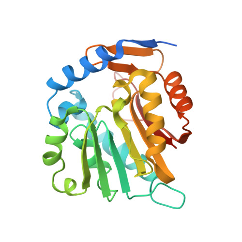 | |
UniProt & NIH Common Fund Data Resources | |||||
GTEx: ENSG00000203740 | |||||
Entity Groups | |||||
| Sequence Clusters | 30% Identity50% Identity70% Identity90% Identity95% Identity100% Identity | ||||
| UniProt Group | Q5VVY1 | ||||
Sequence AnnotationsExpand | |||||
Reference Sequence | |||||
Entity ID: 2 | |||||
|---|---|---|---|---|---|
| Molecule | Chains | Sequence Length | Organism | Details | Image |
| Peptide from Major centromere autoantigen B | C [auth D], D [auth E] | 9 | Homo sapiens | Mutation(s): 0 | 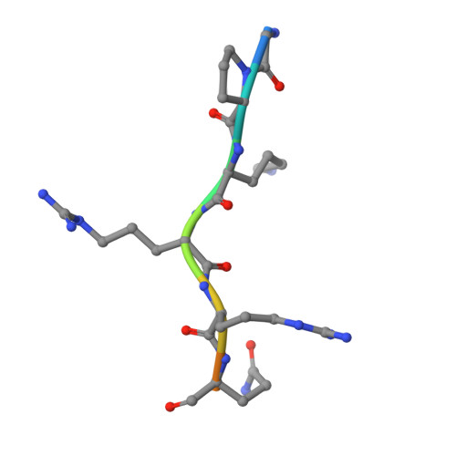 |
UniProt & NIH Common Fund Data Resources | |||||
PHAROS: P07199 GTEx: ENSG00000125817 | |||||
Entity Groups | |||||
| UniProt Group | P07199 | ||||
Sequence AnnotationsExpand | |||||
Reference Sequence | |||||
| Ligands 3 Unique | |||||
|---|---|---|---|---|---|
| ID | Chains | Name / Formula / InChI Key | 2D Diagram | 3D Interactions | |
| SAH (Subject of Investigation/LOI) Download:Ideal Coordinates CCD File | E [auth A], H [auth B] | S-ADENOSYL-L-HOMOCYSTEINE C14 H20 N6 O5 S ZJUKTBDSGOFHSH-WFMPWKQPSA-N |  | ||
| GOL (Subject of Investigation/LOI) Download:Ideal Coordinates CCD File | F [auth A], I [auth B], J [auth B] | GLYCEROL C3 H8 O3 PEDCQBHIVMGVHV-UHFFFAOYSA-N |  | ||
| CL (Subject of Investigation/LOI) Download:Ideal Coordinates CCD File | G [auth A], K [auth B], L [auth E] | CHLORIDE ION Cl VEXZGXHMUGYJMC-UHFFFAOYSA-M |  | ||
| Length ( Å ) | Angle ( ˚ ) |
|---|---|
| a = 70.548 | α = 90 |
| b = 96.534 | β = 90 |
| c = 100.213 | γ = 90 |
| Software Name | Purpose |
|---|---|
| DENZO | data reduction |
| HKL-2000 | data scaling |
| PHENIX | refinement |
| PDB_EXTRACT | data extraction |
| MOLREP | phasing |


