6KAL | pdb_00006kal

Crystal structure of FKRP in complex with Mg ion and CMP

  • Classification: TRANSFERASE
  • Organism(s): Homo sapiens
  • Expression System: Homo sapiens
  • Mutation(s): No 

  • Deposited: 2019-06-23 Released: 2020-01-15 
  • Deposition Author(s): Kuwabara, N.
  • Funding Organization(s): Japan Society for the Promotion of Science

Experimental Data Snapshot

  • Method: X-RAY DIFFRACTION
  • Resolution: 2.60 Å
  • R-Value Free: 
    0.241 (Depositor), 0.242 (DCC) 
  • R-Value Work: 
    0.195 (Depositor), 0.197 (DCC) 
  • R-Value Observed: 
    0.197 (Depositor) 

wwPDB Validation 3D Report Full Report

Validation slider image for 6KAL

Ligand Structure Quality Assessment 


This is version 1.3 of the entry. See complete history

Literature

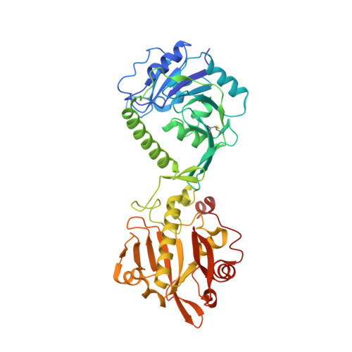
Crystal structures of fukutin-related protein (FKRP), a ribitol-phosphate transferase related to muscular dystrophy.

Kuwabara, N.Imae, R.Manya, H.Tanaka, T.Mizuno, M.Tsumoto, H.Kanagawa, M.Kobayashi, K.Toda, T.Senda, T.Endo, T.Kato, R.

(2020) Nat Commun 11: 303-303

  • DOI: https://doi.org/10.1038/s41467-019-14220-z
  • Primary Citation Related Structures: 
    6KAJ, 6KAK, 6KAL, 6KAM, 6KAN, 6L7S, 6L7T, 6L7U

  • PubMed Abstract: 

    α-Dystroglycan (α-DG) is a highly-glycosylated surface membrane protein. Defects in the O-mannosyl glycan of α-DG cause dystroglycanopathy, a group of congenital muscular dystrophies. The core M3 O-mannosyl glycan contains tandem ribitol-phosphate (RboP), a characteristic feature first found in mammals. Fukutin and fukutin-related protein (FKRP), whose mutated genes underlie dystroglycanopathy, sequentially transfer RboP from cytidine diphosphate-ribitol (CDP-Rbo) to form a tandem RboP unit in the core M3 glycan. Here, we report a series of crystal structures of FKRP with and without donor (CDP-Rbo) and/or acceptor [RboP-(phospho-)core M3 peptide] substrates. FKRP has N-terminal stem and C-terminal catalytic domains, and forms a tetramer both in crystal and in solution. In the acceptor complex, the phosphate group of RboP is recognized by the catalytic domain of one subunit, and a phosphate group on O-mannose is recognized by the stem domain of another subunit. Structure-based functional studies confirmed that the dimeric structure is essential for FKRP enzymatic activity.


  • Organizational Affiliation
    • Structural Biology Research Center, Institute of Materials Structure Science, High Energy Accelerator Research Organization, Tsukuba, Ibaraki, 305-0801, Japan.

Macromolecule Content 

  • Total Structure Weight: 203.4 kDa 
  • Atom Count: 14,174 
  • Modeled Residue Count: 1,772 
  • Deposited Residue Count: 1,820 
  • Unique protein chains: 1

Macromolecules

Find similar proteins by:|  3D Structure
Entity ID: 1
MoleculeChains  Sequence LengthOrganismDetailsImage
Fukutin-related protein
A, B, C, D
455Homo sapiensMutation(s): 0 
Gene Names: FKRP
EC: 2.7.8
UniProt & NIH Common Fund Data Resources
Find proteins for Q9H9S5 (Homo sapiens)
Explore Q9H9S5 
Go to UniProtKB:  Q9H9S5
PHAROS:  Q9H9S5
GTEx:  ENSG00000181027 
Entity Groups
Sequence Clusters30% Identity50% Identity70% Identity90% Identity95% Identity100% Identity
UniProt GroupQ9H9S5
Glycosylation
Glycosylation Sites: 2Go to GlyGen: Q9H9S5-1
Sequence Annotations
Expand
Reference Sequence

Small Molecules

Ligands 4 Unique
IDChains Name / Formula / InChI Key2D Diagram3D Interactions
C5P
(Subject of Investigation/LOI)

Query on C5P


Download:Ideal Coordinates CCD File
G [auth A],
M [auth B],
S [auth C],
Y [auth D]
CYTIDINE-5'-MONOPHOSPHATE
C9 H14 N3 O8 P
IERHLVCPSMICTF-XVFCMESISA-N
NAG

Query on NAG


Download:Ideal Coordinates CCD File
F [auth A]
K [auth B]
L [auth B]
Q [auth C]
R [auth C]
F [auth A],
K [auth B],
L [auth B],
Q [auth C],
R [auth C],
W [auth D],
X [auth D]
2-acetamido-2-deoxy-beta-D-glucopyranose
C8 H15 N O6
OVRNDRQMDRJTHS-FMDGEEDCSA-N
ZN
(Subject of Investigation/LOI)

Query on ZN


Download:Ideal Coordinates CCD File
E [auth A],
J [auth B],
P [auth C],
V [auth D]
ZINC ION
Zn
PTFCDOFLOPIGGS-UHFFFAOYSA-N
MG
(Subject of Investigation/LOI)

Query on MG


Download:Ideal Coordinates CCD File
AA [auth D]
H [auth A]
I [auth A]
N [auth B]
O [auth B]
AA [auth D],
H [auth A],
I [auth A],
N [auth B],
O [auth B],
T [auth C],
U [auth C],
Z [auth D]
MAGNESIUM ION
Mg
JLVVSXFLKOJNIY-UHFFFAOYSA-N

Experimental Data & Validation

Experimental Data

  • Method: X-RAY DIFFRACTION
  • Resolution: 2.60 Å
  • R-Value Free:  0.241 (Depositor), 0.242 (DCC) 
  • R-Value Work:  0.195 (Depositor), 0.197 (DCC) 
  • R-Value Observed: 0.197 (Depositor) 
Space Group: P 21 21 21
Unit Cell:
Length ( Å )Angle ( ˚ )
a = 77.583α = 90
b = 118.674β = 90
c = 252.695γ = 90
Software Package:
Software NamePurpose
PHENIXrefinement
XDSdata reduction
Aimlessdata scaling
PHASERphasing

Structure Validation

View Full Validation Report


Ligand Structure Quality Assessment 


Entry History 

& Funding Information

Deposition Data

  • Released Date: 2020-01-15 
  • Deposition Author(s): Kuwabara, N.

Funding OrganizationLocationGrant Number
Japan Society for the Promotion of ScienceJapan16K07284
Japan Society for the Promotion of ScienceJapan26840029

Revision History  (Full details and data files)

  • Version 1.0: 2020-01-15
    Type: Initial release
  • Version 1.1: 2020-01-29
    Changes: Database references
  • Version 1.2: 2020-07-29
    Type: Remediation
    Reason: Carbohydrate remediation
    Changes: Data collection, Derived calculations, Structure summary
  • Version 1.3: 2024-11-06
    Changes: Data collection, Database references, Derived calculations, Structure summary