6K96 | pdb_00006k96

Crystal structure of Ari2


Experimental Data Snapshot

  • Method: X-RAY DIFFRACTION
  • Resolution: 2.50 Å
  • R-Value Free: 
    0.280 (Depositor), 0.279 (DCC) 
  • R-Value Work: 
    0.226 (Depositor), 0.229 (DCC) 
  • R-Value Observed: 
    0.229 (Depositor) 

Starting Model: experimental
View more details

wwPDB Validation 3D Report Full Report

Validation slider image for 6K96

Ligand Structure Quality Assessment 


This is version 1.2 of the entry. See complete history

Literature

Stereochemistry in the Reaction of themyo-Inositol Phosphate Synthase Ortholog Ari2 during Aristeromycin Biosynthesis.

Kudo, F.Tsunoda, T.Yamaguchi, K.Miyanaga, A.Eguchi, T.

(2019) Biochemistry 58: 5112-5116

  • DOI: https://doi.org/10.1021/acs.biochem.9b00981
  • Primary Citation Related Structures: 
    6K96

  • PubMed Abstract: 

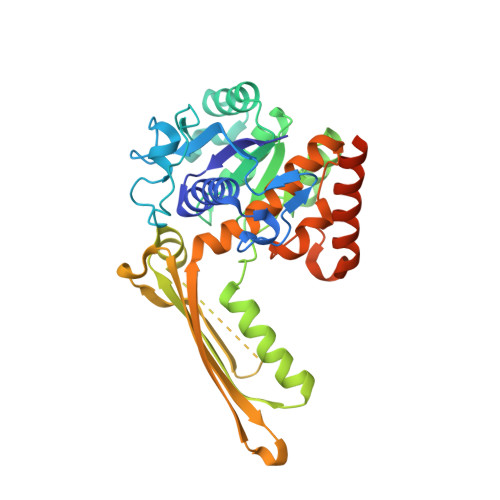
    The myo -inositol-1-phosphate synthase (MIPS) ortholog Ari2, which is encoded in the aristeromycin biosynthetic gene cluster, catalyzes the formation of five-membered cyclitol phosphate using d-fructose 6-phosphate (F6P) as a substrate. To understand the stereochemistry during the Ari2 reaction in vivo , we carried out feeding experiments with (6 S )-d-[6- 2 H 1 ]- and (6 R )-d-[6- 2 H 1 ]glucose in the aristeromycin-producing strain Streptomyces citricolor . We observed retention of the 2 H atom of (6 S )-d-[6- 2 H 1 ]glucose and no incorporation of the 2 H atom from (6 R )-d-[6- 2 H 1 ]glucose in aristeromycin. This indicates that Ari2 abstracts the pro-R proton at C6 of F6P after oxidation of C5-OH by nicotinamide adenine dinucleotide (NAD + ) to generate the enolate intermediate, which then attacks the C2 ketone to form the C-C bond via aldol-type condensation. The reaction of Ari2 with (6 S )-d-[6- 2 H 1 ]- and (6 R )-d-[6- 2 H 1 ]F6P in vitro exhibited identical stereochemistry compared with that observed during the feeding experiments. Furthermore, analysis of the crystal structure of Ari2, including NAD + as a ligand, revealed the active site of Ari2 to be similar to that of MIPS of Mycobacterium tuberculosis , supporting the similarity of the reaction mechanisms of Ari2 and MIPS.


  • Organizational Affiliation
    • Department of Chemistry , Tokyo Institute of Technology , 2-12-1, O-okayama , Meguro-ku, Tokyo 152-8551 , Japan.

Macromolecule Content 

  • Total Structure Weight: 82.07 kDa 
  • Atom Count: 5,365 
  • Modeled Residue Count: 676 
  • Deposited Residue Count: 756 
  • Unique protein chains: 1

Macromolecules

Find similar proteins by:|  3D Structure
Entity ID: 1
MoleculeChains  Sequence LengthOrganismDetailsImage
Five-membered-cyclitol-phosphate synthase
A, B
378Streptomyces citricolorMutation(s): 0 
Gene Names: ari2
UniProt
Find proteins for A0A1B4ZC85 (Streptomyces citricolor)
Explore A0A1B4ZC85 
Go to UniProtKB:  A0A1B4ZC85
Entity Groups
Sequence Clusters30% Identity50% Identity70% Identity90% Identity95% Identity100% Identity
UniProt GroupA0A1B4ZC85
Sequence Annotations
Expand
Reference Sequence

Small Molecules

Ligands 3 Unique
IDChains Name / Formula / InChI Key2D Diagram3D Interactions
NAD
(Subject of Investigation/LOI)

Query on NAD


Download:Ideal Coordinates CCD File
C [auth A],
G [auth B]
NICOTINAMIDE-ADENINE-DINUCLEOTIDE
C21 H27 N7 O14 P2
BAWFJGJZGIEFAR-NNYOXOHSSA-N
GOL

Query on GOL


Download:Ideal Coordinates CCD File
E [auth A],
F [auth A],
I [auth B]
GLYCEROL
C3 H8 O3
PEDCQBHIVMGVHV-UHFFFAOYSA-N
NA

Query on NA


Download:Ideal Coordinates CCD File
D [auth A],
H [auth B]
SODIUM ION
Na
FKNQFGJONOIPTF-UHFFFAOYSA-N

Experimental Data & Validation

Experimental Data

  • Method: X-RAY DIFFRACTION
  • Resolution: 2.50 Å
  • R-Value Free:  0.280 (Depositor), 0.279 (DCC) 
  • R-Value Work:  0.226 (Depositor), 0.229 (DCC) 
  • R-Value Observed: 0.229 (Depositor) 
Space Group: P 65 2 2
Unit Cell:
Length ( Å )Angle ( ˚ )
a = 101.18α = 90
b = 101.18β = 90
c = 389.662γ = 120
Software Package:
Software NamePurpose
REFMACrefinement
iMOSFLMdata reduction
SCALAdata scaling
MOLREPphasing

Structure Validation

View Full Validation Report


Ligand Structure Quality Assessment 


Entry History 

Deposition Data

Revision History  (Full details and data files)

  • Version 1.0: 2019-12-25
    Type: Initial release
  • Version 1.1: 2020-01-08
    Changes: Database references
  • Version 1.2: 2023-11-22
    Changes: Data collection, Database references, Derived calculations, Refinement description