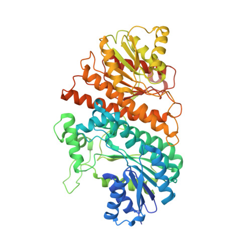
6K8V

Crystal structure of N-domain of baterial malonyl-CoA reductase


Experimental Data Snapshot

  • Method: X-RAY DIFFRACTION
  • Resolution: 2.20 Å
  • R-Value Free: 0.258 
  • R-Value Work: 0.198 
  • R-Value Observed: 0.201 

wwPDB Validation   3D Report Full Report


Ligand Structure Quality Assessment 


This is version 1.1 of the entry. See complete history


Literature

Structural insight into bi-functional malonyl-CoA reductase.

Son, H.F.Kim, S.Seo, H.Hong, J.Lee, D.Jin, K.S.Park, S.Kim, K.J.

(2020) Environ Microbiol 22: 752-765

  • DOI: https://doi.org/10.1111/1462-2920.14885
  • Primary Citation of Related Structures:  
    6K8S, 6K8T, 6K8U, 6K8V, 6K8W

  • PubMed Abstract: 

    The bi-functional malonyl-CoA reductase is a key enzyme of the 3-hydroxypropionate bi-cycle for bacterial CO 2 fixation, catalysing the reduction of malonyl-CoA to malonate semialdehyde and further reduction to 3-hydroxypropionate. Here, we report the crystal structure and the full-length architecture of malonyl-CoA reductase from Porphyrobacter dokdonensis. The malonyl-CoA reductase monomer of 1230 amino acids consists of four tandemly arranged short-chain dehydrogenases/reductases, with two catalytic and two non-catalytic short-chain dehydrogenases/reductases, and forms a homodimer through paring contact of two malonyl-CoA reductase monomers. The complex structures with its cofactors and substrates revealed that the malonyl-CoA substrate site is formed by the cooperation of two short-chain dehydrogenases/reductases and one novel extra domain, while only one catalytic short-chain dehydrogenase/reductase contributes to the formation of the malonic semialdehyde-binding site. The phylogenetic and structural analyses also suggest that the bacterial bi-functional malonyl-CoA has a structural origin that is completely different from the archaeal mono-functional malonyl-CoA and malonic semialdehyde reductase, and thereby constitute an efficient enzyme.


  • Organizational Affiliation

    School of Life Sciences (KNU Creative BioResearch Group), KNU Institute for Microorganisms, Kyungpook National University, Daegu, South Korea.


Macromolecules
Find similar proteins by:  (by identity cutoff)  |  3D Structure
Entity ID: 1
MoleculeChains Sequence LengthOrganismDetailsImage
NAD-dependent epimerase/dehydratase:Short-chain dehydrogenase/reductase SDR
A, B
575Erythrobacter dokdonensis DSW-74Mutation(s): 0 
Gene Names: I603_0811
UniProt
Find proteins for A0A1A7BFR5 (Erythrobacter dokdonensis DSW-74)
Explore A0A1A7BFR5 
Go to UniProtKB:  A0A1A7BFR5
Entity Groups  
Sequence Clusters30% Identity50% Identity70% Identity90% Identity95% Identity100% Identity
UniProt GroupA0A1A7BFR5
Sequence Annotations
Expand
  • Reference Sequence
Small Molecules
Ligands 2 Unique
IDChains Name / Formula / InChI Key2D Diagram3D Interactions
SO4 (Subject of Investigation/LOI)
Query on SO4

Download Ideal Coordinates CCD File 
C [auth A]
D [auth A]
E [auth A]
F [auth A]
G [auth A]
C [auth A],
D [auth A],
E [auth A],
F [auth A],
G [auth A],
H [auth A],
I [auth A],
K [auth B],
L [auth B],
M [auth B],
N [auth B],
O [auth B],
P [auth B]
SULFATE ION
O4 S
QAOWNCQODCNURD-UHFFFAOYSA-L
GOL (Subject of Investigation/LOI)
Query on GOL

Download Ideal Coordinates CCD File 
J [auth B]GLYCEROL
C3 H8 O3
PEDCQBHIVMGVHV-UHFFFAOYSA-N
Experimental Data & Validation

Experimental Data

  • Method: X-RAY DIFFRACTION
  • Resolution: 2.20 Å
  • R-Value Free: 0.258 
  • R-Value Work: 0.198 
  • R-Value Observed: 0.201 
  • Space Group: P 21 21 21
Unit Cell:
Length ( Å )Angle ( ˚ )
a = 85.637α = 90
b = 109.072β = 90
c = 129.422γ = 90
Software Package:
Software NamePurpose
HKL-2000data reduction
REFMACrefinement
PDB_EXTRACTdata extraction
HKL-2000data scaling
PHENIXphasing

Structure Validation

View Full Validation Report


Ligand Structure Quality Assessment 


Entry History 

Deposition Data

Revision History  (Full details and data files)

  • Version 1.0: 2020-03-18
    Type: Initial release
  • Version 1.1: 2024-03-27
    Changes: Data collection, Database references