6K8G | pdb_00006k8g

H/D exchanged Hen egg-white lysozyme

  • Classification: HYDROLASE
  • Organism(s): Gallus gallus
  • Mutation(s): No 

  • Deposited: 2019-06-11 Released: 2020-06-17 
  • Deposition Author(s): Kita, A., Morimoto, Y.
  • Funding Organization(s): Ministry of Education, Culture, Sports, Science and Technology (Japan), Other private

Experimental Data Snapshot

  • Method: X-RAY DIFFRACTION
  • Resolution: 2.00 Å
  • R-Value Free: 
    0.180 (Depositor), 0.186 (DCC) 
  • R-Value Work: 
    0.132 (Depositor), 0.135 (DCC) 
  • R-Value Observed: 
    0.134 (Depositor) 

  • Method: NEUTRON DIFFRACTION
  • Resolution: 2.00 Å
  • R-Value Free: 
    0.255 (Depositor) 
  • R-Value Work: 
    0.225 (Depositor) 

Starting Model: experimental
View more details

wwPDB Validation 3D Report Full Report

Validation slider image for 6K8G

This is version 1.3 of the entry. See complete history

Literature

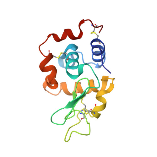
Hydrogen/deuterium exchange behavior in tetragonal hen egg-white lysozyme crystals affected by solution state.

Kita, A.Morimoto, Y.

(2020) J Appl Crystallogr 53: 837-840

  • DOI: https://doi.org/10.1107/S1600576720005488
  • Primary Citation Related Structures: 
    6K8G

  • PubMed Abstract: 

    Neutron diffraction studies of hydrogen/deuterium-exchanged hen egg-white lysozyme were performed by a joint X-ray and neutron refinement to elucidate the hydrogen/deuterium exchange behavior. Large crystals for neutron work, consisting of molecules that were exchanged before crystallization, were obtained by repeatedly adding protein solution to the crystal batch using deuterated precipitant reagent. There are differences in hydrogen/deuterium exchange behavior compared with previous crystallographic or NMR studies, which could be due to intermolecular interactions in the crystal or to different lengths of exchange period.


  • Organizational Affiliation
    • Institute for Integrated Radiation and Nuclear Science, Kyoto University, Kumatori, Sen-nan, Osaka 590-0494, Japan.

Macromolecule Content 

  • Total Structure Weight: 14.39 kDa 
  • Atom Count: 1,086 
  • Modeled Residue Count: 129 
  • Deposited Residue Count: 129 
  • Unique protein chains: 1

Macromolecules

Find similar proteins by:|  3D Structure
Entity ID: 1
MoleculeChains  Sequence LengthOrganismDetailsImage
Lysozyme C129Gallus gallusMutation(s): 0 
EC: 3.2.1.17
UniProt
Find proteins for P00698 (Gallus gallus)
Explore P00698 
Go to UniProtKB:  P00698
Entity Groups
Sequence Clusters30% Identity50% Identity70% Identity90% Identity95% Identity100% Identity
UniProt GroupP00698
Sequence Annotations
Expand
Reference Sequence

Small Molecules

Experimental Data & Validation

Experimental Data

  • Method: X-RAY DIFFRACTION
  • Resolution: 2.00 Å
  • R-Value Free:  0.180 (Depositor), 0.186 (DCC) 
  • R-Value Work:  0.132 (Depositor), 0.135 (DCC) 
  • R-Value Observed: 0.134 (Depositor) 
  • Method: NEUTRON DIFFRACTION
  • Resolution: 2.00 Å
  • R-Value Free:  0.255 (Depositor) 
  • R-Value Work:  0.225 (Depositor) 
Space Group: P 43 21 2
Unit Cell:
Length ( Å )Angle ( ˚ )
a = 79.094α = 90
b = 79.094β = 90
c = 37.828γ = 90
Software Package:
Software NamePurpose
PHENIXrefinement
PHENIXrefinement
HKL-2000data reduction
HKL-2000data scaling
MOLREPphasing

Structure Validation

View Full Validation Report


Entry History 

& Funding Information

Deposition Data


Funding OrganizationLocationGrant Number
Ministry of Education, Culture, Sports, Science and Technology (Japan)Japan--
Other privateJapanThe Research Development Program of Kyoto University
Other privateJapanThe Towa Foundation for Food Science & Research

Revision History  (Full details and data files)

  • Version 1.0: 2020-06-17
    Type: Initial release
  • Version 1.1: 2021-01-13
    Changes: Database references
  • Version 1.2: 2024-04-03
    Changes: Data collection, Database references, Refinement description
  • Version 1.3: 2024-11-20
    Changes: Structure summary