6K2O | pdb_00006k2o

Structural basis of glycan recognition in globally predominant human P[8] rotavirus


Experimental Data Snapshot

  • Method: X-RAY DIFFRACTION
  • Resolution: 2.30 Å
  • R-Value Free: 
    0.233 (Depositor), 0.234 (DCC) 
  • R-Value Work: 
    0.203 (Depositor), 0.209 (DCC) 
  • R-Value Observed: 
    0.205 (Depositor) 

Starting Model: experimental
View more details

wwPDB Validation 3D Report Full Report

Validation slider image for 6K2O

This is version 2.1 of the entry. See complete history

Literature

Structural Basis of Glycan Recognition in Globally Predominant Human P[8] Rotavirus.

Sun, X.Dang, L.Li, D.Qi, J.Wang, M.Chai, W.Zhang, Q.Wang, H.Bai, R.Tan, M.Duan, Z.

(2020) Virol Sin 35: 156-170

  • DOI: https://doi.org/10.1007/s12250-019-00164-7
  • Primary Citation Related Structures: 
    6K2N, 6K2O

  • PubMed Abstract: 

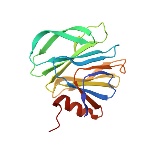
    Rotavirus (RV) causes acute gastroenteritis in infants and children worldwide. Recent studies showed that glycans such as histo-blood group antigens (HBGAs) function as cell attachment factors affecting RV host susceptibility and prevalence. P[8] is the predominant RV genotype in humans, but the structural basis of how P[8] RVs interact with glycan ligands remains elusive. In this study, we characterized the interactions between P[8] VP8*s and glycans which showed that VP8*, the RV glycan binding domain, recognized both mucin core 2 and H type 1 antigens according to the ELISA-based oligosaccharide binding assays. Importantly, we determined the structural basis of P[8] RV-glycans interaction from the crystal structures of a Rotateq P[8] VP8* in complex with core 2 and H type 1 glycans at 1.8 Å and 2.3 Å, respectively, revealing a common binding pocket and similar binding mode. Structural and sequence analysis demonstrated that the glycan binding site is conserved among RVs in the P[II] genogroup, while genotype-specific amino acid variations determined different glycan binding preference. Our data elucidated the detailed structural basis of the interactions between human P[8] RVs and different host glycan factors, shedding light on RV infection, epidemiology, and development of anti-viral agents.


  • Organizational Affiliation
    • National Health Commission Key Laboratory for Medical Virology and Viral Diseases, Beijing, 102206, China.

Macromolecule Content 

  • Total Structure Weight: 19.61 kDa 
  • Atom Count: 1,463 
  • Modeled Residue Count: 163 
  • Deposited Residue Count: 163 
  • Unique protein chains: 1

Macromolecules

Find similar proteins by:|  3D Structure
Entity ID: 1
MoleculeChains  Sequence LengthOrganismDetailsImage
Outer capsid protein VP4163Rotavirus AMutation(s): 0 
UniProt
Find proteins for E2EA82 (Rotavirus A)
Explore E2EA82 
Go to UniProtKB:  E2EA82
Entity Groups
Sequence Clusters30% Identity50% Identity70% Identity90% Identity95% Identity100% Identity
UniProt GroupE2EA82
Sequence Annotations
Expand
Reference Sequence

Oligosaccharides

Help  
Entity ID: 2
MoleculeChains Length2D Diagram GlycosylationD Interactions
alpha-L-fucopyranose-(1-2)-beta-D-galactopyranose-(1-3)-2-acetamido-2-deoxy-beta-D-glucopyranose-(1-3)-beta-D-galactopyranose
B
4N/A
Glycosylation Resources
GlyTouCan: G00294TN
GlyCosmos: G00294TN
GlyGen: G00294TN

Small Molecules

Experimental Data & Validation

Experimental Data

  • Method: X-RAY DIFFRACTION
  • Resolution: 2.30 Å
  • R-Value Free:  0.233 (Depositor), 0.234 (DCC) 
  • R-Value Work:  0.203 (Depositor), 0.209 (DCC) 
  • R-Value Observed: 0.205 (Depositor) 
Space Group: P 63 2 2
Unit Cell:
Length ( Å )Angle ( ˚ )
a = 80.454α = 90
b = 80.454β = 90
c = 116.593γ = 120
Software Package:
Software NamePurpose
HKL-2000data reduction
HKL-2000data scaling
PHENIXrefinement
PDB_EXTRACTdata extraction
PHASERphasing

Structure Validation

View Full Validation Report


Entry History 

& Funding Information

Deposition Data


Funding OrganizationLocationGrant Number
National Science Foundation (China)Chinano.2018ZA10711-001 and no.2017ZX10104001

Revision History  (Full details and data files)

  • Version 1.0: 2019-10-09
    Type: Initial release
  • Version 1.1: 2019-10-30
    Changes: Data collection, Database references
  • Version 1.2: 2020-05-13
    Changes: Data collection, Database references
  • Version 2.0: 2020-07-29
    Type: Remediation
    Reason: Carbohydrate remediation
    Changes: Advisory, Atomic model, Data collection, Derived calculations, Structure summary
  • Version 2.1: 2023-11-22
    Changes: Data collection, Database references, Derived calculations, Refinement description, Structure summary