6JZ8 | pdb_00006jz8

b-glucuronidase from Ruminococcus gnavus in complex with D-glucaro 1,5-lactone


Experimental Data Snapshot

  • Method: X-RAY DIFFRACTION
  • Resolution: 1.58 Å
  • R-Value Free: 
    0.174 (Depositor) 
  • R-Value Work: 
    0.163 (Depositor) 

Starting Model: experimental
View more details

wwPDB Validation 3D Report Full Report

Validation slider image for 6JZ8

Ligand Structure Quality Assessment 


This is version 2.2 of the entry. See complete history

Literature

Substituent Position of Iminocyclitols Determines the Potency and Selectivity for Gut Microbial Xenobiotic-Reactivating Enzymes.

Dashnyam, P.Lin, H.Y.Chen, C.Y.Gao, S.Yeh, L.F.Hsieh, W.C.Tu, Z.Lin, C.H.

(2020) J Med Chem 63: 4617-4627

  • DOI: https://doi.org/10.1021/acs.jmedchem.9b01918
  • Primary Citation Related Structures: 
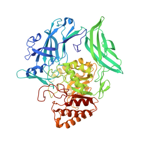
    6JZ1, 6JZ2, 6JZ3, 6JZ4, 6JZ5, 6JZ6, 6JZ7, 6JZ8

  • PubMed Abstract: 

    Selective inhibitors of gut bacterial β-glucuronidases (GUSs) are of particular interest in the prevention of xenobiotic-induced toxicities. This study reports the first structure-activity relationships on potency and selectivity of several iminocyclitols ( 2 - 7 ) for the GUSs. Complex structures of Ruminococcus gnavus GUS with 2 - 7 explained how charge, conformation, and substituent of iminocyclitols affect their potency and selectivity. N1 of uronic isofagomine ( 2 ) made strong electrostatic interactions with two catalytic glutamates of GUSs, resulting in the most potent inhibition ( K i ≥ 11 nM). C6-propyl analogue of 2 ( 6 ) displayed 700-fold selectivity for opportunistic bacterial GUSs ( K i = 74 nM for E. coli GUS and 51.8 μM for Rg GUS). In comparison with 2 , there was 200-fold enhancement in the selectivity, which was attributed to differential interactions between the propyl group and loop 5 residues of the GUSs. The results provide useful insights to develop potent and selective inhibitors for undesired GUSs.


  • Organizational Affiliation
    • Institute of Biological Chemistry, Academia Sinica, No 128, Academia Road, Taipei 11529, Taiwan.

Macromolecule Content 

  • Total Structure Weight: 140.46 kDa 
  • Atom Count: 10,614 
  • Modeled Residue Count: 1,158 
  • Deposited Residue Count: 1,206 
  • Unique protein chains: 1

Macromolecules

Find similar proteins by:|  3D Structure
Entity ID: 1
MoleculeChains  Sequence LengthOrganismDetailsImage
Beta-glucuronidase
A, B
603Mediterraneibacter gnavusMutation(s): 0 
Gene Names: uidA
EC: 3.2.1.31
UniProt
Find proteins for Q6W7J7 (Mediterraneibacter gnavus)
Explore Q6W7J7 
Go to UniProtKB:  Q6W7J7
Entity Groups
Sequence Clusters30% Identity50% Identity70% Identity90% Identity95% Identity100% Identity
UniProt GroupQ6W7J7
Sequence Annotations
Expand
Reference Sequence

Small Molecules

Experimental Data & Validation

Experimental Data

  • Method: X-RAY DIFFRACTION
  • Resolution: 1.58 Å
  • R-Value Free:  0.174 (Depositor) 
  • R-Value Work:  0.163 (Depositor) 
Space Group: C 1 2 1
Unit Cell:
Length ( Å )Angle ( ˚ )
a = 162.56α = 90
b = 103.345β = 130.99
c = 112.281γ = 90
Software Package:
Software NamePurpose
PHENIXrefinement
HKL-2000data scaling
PDB_EXTRACTdata extraction
HKL-2000data reduction
PHENIXphasing

Structure Validation

View Full Validation Report


Ligand Structure Quality Assessment 


Entry History 

& Funding Information

Deposition Data


Funding OrganizationLocationGrant Number
Ministry of Science and Technology (MoST, Taiwan)Taiwan--

Revision History  (Full details and data files)

  • Version 1.0: 2020-06-10
    Type: Initial release
  • Version 2.0: 2020-07-29
    Type: Remediation
    Reason: Carbohydrate remediation
    Changes: Atomic model, Derived calculations, Structure summary
  • Version 2.1: 2020-12-23
    Changes: Database references
  • Version 2.2: 2023-11-22
    Changes: Data collection, Database references, Refinement description