GII.13/21 Noroviruses Recognize Glycans with a Terminal beta-Galactose via an Unconventional Glycan Binding Site.
Cong, X., Sun, X.M., Qi, J.X., Li, H.B., Chai, W.G., Zhang, Q., Wang, H., Kong, X.Y., Song, J., Pang, L.L., Jin, M., Li, D.D., Tan, M., Duan, Z.J.(2019) J Virol 93
- PubMed: 31118252
- DOI: https://doi.org/10.1128/JVI.00723-19
- Primary Citation of Related Structures:
6JYN, 6JYO, 6JYR, 6JYS - PubMed Abstract:
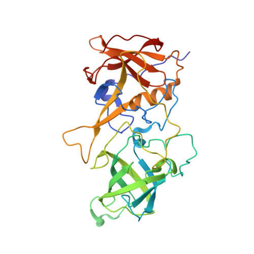
Human noroviruses (huNoVs) recognize histo-blood group antigens (HBGAs) as host susceptibility factors. GII.13 and GII.21 huNoVs form a unique genetic lineage that emerged from mainstream GII NoVs via development of a new, nonconventional glycan binding site (GBS) that binds Le a antigen. This previous finding raised the question of whether the new GII.13/21 GBS really has such a narrow glycan binding spectrum. In this study, we provide solid phenotypic and structural evidence indicating that this new GBS recognizes a group of glycans with a common terminal β-galactose (β-Gal). First, we found that P domain proteins of GII.13/21 huNoVs circulating at different times bound three glycans sharing a common terminal β-Gal, including Lec, lactose, and mucin core 2. Second, we solved the crystal structures of the GII.13 P dimers in complex with Lec and mucin core 2, which showed that β-Gal is the major binding saccharide. Third, nonfat milk and lactose blocked the GII.13/21 P domain-glycan binding, which may explain the low prevalence of GII.13/21 viruses. Our data provide new insight into the host interactions and epidemiology of huNoVs, which would help in the control and prevention of NoV-associated diseases. IMPORTANCE Evidence from both phenotypic binding assay and structural study support the observed interactions of human noroviruses (huNoVs) with histo-blood group antigens (HBGAs) as receptors or attachment factors, affecting their host susceptibility. GII.13 and GII.21 genotypes form a unique genetic lineage that differs from the mainstream GII huNoVs in their unconventional glycan binding site. Unlike the previous findings that GII.13/21 genotypes recognize only Le a antigen, we found in this study that they can interact with a group of glycans with a common terminal β-Gal, including Lec, lactose, and mucin core 2. However, this wide glycan binding spectrum in a unique binding mode of the GII.13/21 huNoVs appears not to increase their prevalence, probably due to the existence of decoy glycan receptors in human gastrointestinal tract limiting their infection. Our findings shed light on the host interaction and epidemiology of huNoVs, which would impact the strategy of huNoV control and prevention.
Organizational Affiliation:
Key Laboratory for Medical Virology and Viral Diseases, National Health Commission of the People's Republic of China, Beijing, China.


