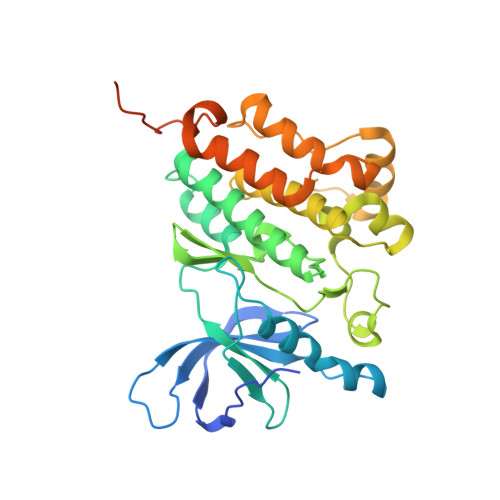
6JX0

Crystal structure of EGFR 696-1022 T790M in complex with AZD9291 prepared by co-crystallization


Experimental Data Snapshot

  • Method: X-RAY DIFFRACTION
  • Resolution: 2.53 Å
  • R-Value Free: 0.222 
  • R-Value Work: 0.192 
  • R-Value Observed: 0.193 

wwPDB Validation   3D Report Full Report


Ligand Structure Quality Assessment 


This is version 1.3 of the entry. See complete history


Literature

Structural Basis of AZD9291 Selectivity for EGFR T790M.

Yan, X.E.Ayaz, P.Zhu, S.J.Zhao, P.Liang, L.Zhang, C.H.Wu, Y.C.Li, J.L.Choi, H.G.Huang, X.Shan, Y.Shaw, D.E.Yun, C.H.

(2020) J Med Chem 63: 8502-8511

  • DOI: https://doi.org/10.1021/acs.jmedchem.0c00891
  • Primary Citation of Related Structures:  
    6JWL, 6JX0, 6JX4, 6JXT

  • PubMed Abstract: 

    AZD9291 (Osimertinib) is highly effective in treating EGFR-mutated non-small-cell lung cancers (NSCLCs) with T790M-mediated drug resistance. Despite the remarkable success of AZD9291, its binding pose with EGFR T790M remains unclear. Here, we report unbiased, atomic-level molecular dynamics (MD) simulations in which spontaneous association of AZD9291 with EGFR kinases having WT and T790M mutant gatekeepers was observed. Simulation-generated structural models suggest that the binding pose of AZD9291 with T790M differs from its binding pose with the WT, and that AZD9291 interacts extensively with the gatekeeper residue (Met 790) in T790M but not with Thr 790 in the WT, which explains why AZD9291 binds T790M with higher affinity. The MD simulation-generated models were confirmed by experimentally determined EGFR/T790M complex crystal structures. This work may facilitate the rational design of drugs that can overcome resistance mutations to AZD9291, and more generally it suggests the potential of using unbiased MD simulation to elucidate small-molecule binding poses.


  • Organizational Affiliation

    D. E. Shaw Research, New York, New York 10036, United States.


Macromolecules
Find similar proteins by:  (by identity cutoff)  |  3D Structure
Entity ID: 1
MoleculeChains Sequence LengthOrganismDetailsImage
Epidermal growth factor receptor331Homo sapiensMutation(s): 1 
Gene Names: EGFRERBBERBB1HER1
EC: 2.7.10.1
UniProt & NIH Common Fund Data Resources
Find proteins for P00533 (Homo sapiens)
Explore P00533 
Go to UniProtKB:  P00533
PHAROS:  P00533
GTEx:  ENSG00000146648 
Entity Groups  
Sequence Clusters30% Identity50% Identity70% Identity90% Identity95% Identity100% Identity
UniProt GroupP00533
Sequence Annotations
Expand
  • Reference Sequence
Small Molecules
Binding Affinity Annotations 
IDSourceBinding Affinity
YY3 BindingDB:  6JX0 Ki: min: 2.2, max: 434 (nM) from 6 assay(s)
IC50: min: 2.00e-3, max: 19 (nM) from 55 assay(s)
EC50: min: 6, max: 7930 (nM) from 19 assay(s)
Experimental Data & Validation

Experimental Data

  • Method: X-RAY DIFFRACTION
  • Resolution: 2.53 Å
  • R-Value Free: 0.222 
  • R-Value Work: 0.192 
  • R-Value Observed: 0.193 
  • Space Group: P 31 2 1
Unit Cell:
Length ( Å )Angle ( ˚ )
a = 94.464α = 90
b = 94.464β = 90
c = 125.95γ = 120
Software Package:
Software NamePurpose
PHENIXrefinement
HKL-3000data reduction
HKL-3000data scaling
PHASERphasing

Structure Validation

View Full Validation Report


Ligand Structure Quality Assessment 


Entry History 

Deposition Data

Revision History  (Full details and data files)

  • Version 1.0: 2020-04-22
    Type: Initial release
  • Version 1.1: 2020-09-09
    Changes: Structure summary
  • Version 1.2: 2020-11-04
    Changes: Database references
  • Version 1.3: 2024-10-23
    Changes: Data collection, Database references, Structure summary