6JWV | pdb_00006jwv

Crystal structure of Plasmodium falciparum HPPK-DHPS A437G with STZ-DHP


Experimental Data Snapshot

  • Method: X-RAY DIFFRACTION
  • Resolution: 2.70 Å
  • R-Value Free: 
    0.257 (Depositor), 0.249 (DCC) 
  • R-Value Work: 
    0.206 (Depositor), 0.202 (DCC) 
  • R-Value Observed: 
    0.211 (Depositor) 

Starting Model: experimental
View more details

wwPDB Validation 3D Report Full Report

Validation slider image for 6JWV

Ligand Structure Quality Assessment 


This is version 1.2 of the entry. See complete history

Literature

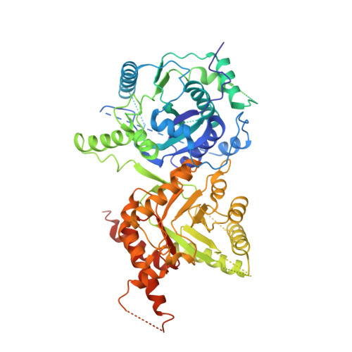
The structure of Plasmodium falciparum hydroxymethyldihydropterin pyrophosphokinase-dihydropteroate synthase reveals the basis of sulfa resistance.

Chitnumsub, P.Jaruwat, A.Talawanich, Y.Noytanom, K.Liwnaree, B.Poen, S.Yuthavong, Y.

(2020) FEBS J 287: 3273-3297

  • DOI: https://doi.org/10.1111/febs.15196
  • Primary Citation Related Structures: 
    6JWQ, 6JWR, 6JWS, 6JWT, 6JWU, 6JWV, 6JWW, 6JWX, 6JWY, 6JWZ, 6KCK, 6KCL, 6KCM

  • PubMed Abstract: 

    The clinical efficacy of sulfa drugs as antimalarials has declined owing to the evolution of resistance in Plasmodium falciparum (Pf) malaria parasites. In order to understand the basis of this resistance and to design more effective antimalarials, we have solved 13 structures of the bifunctional enzyme 6-hydroxymethyl-7,8-dihydropterin pyrophosphokinase (HPPK)-dihydropteroate synthase (DHPS) from wild-type (WT) P. falciparum and sulfa-resistant mutants, both as apoenzyme and as complexes with pteroate (PTA) and sulfa derivatives. The structures of these complexes show that PTA, which effectively inhibits both the WT and mutants, stays in active sites without steric constraint. In contrast, parts of the sulfa compounds situated outside of the substrate envelope are in the vicinity of the resistance mutations. Steric conflict between compound and mutant residue along with increased flexibility of loop D2 in the mutants can account for the reduced compound binding affinity to the mutants. Kinetic data show that the mutants have enhanced enzyme activity compared with the WT. These PfDHPS structural insights are critical for the design of novel, substrate envelope-compliant DHPS inhibitors that are less vulnerable to resistance mutations. DATABASES: The data reported in this paper have been deposited in the Protein Data Bank, www.wwpdb.org. PDB ID codes: 6JWQ for apoWT; 6JWR, 6JWS, and 6JWT for PTA complexes of WT, A437G (3D7), and V1/S; 6JWU, 6JWV, and 6JWW for STZ-DHP complexes of WT, 3D7, and V1/S; 6JWX, 6JWY, and 6JWZ for SDX-DHP complexes of WT, 3D7, and W2; 6KCK, 6KCL, and 6KCM for Pterin/pHBA complexes of WT, TN1, and W2.


  • Organizational Affiliation
    • National Center for Genetic Engineering and Biotechnology (BIOTEC), National Science and Technology Development Agency, Pathumthani, Thailand.

Macromolecule Content 

  • Total Structure Weight: 174.03 kDa 
  • Atom Count: 9,403 
  • Modeled Residue Count: 1,083 
  • Deposited Residue Count: 1,456 
  • Unique protein chains: 1

Macromolecules

Find similar proteins by:|  3D Structure
Entity ID: 1
MoleculeChains  Sequence LengthOrganismDetailsImage
7,8-dihydro-6-hydroxymethylpterin pyrophosphokinase-dihydropteroate synthase
A, B
728Plasmodium falciparumMutation(s): 1 
Gene Names: PPPK-DHPS
UniProt
Find proteins for Q25704 (Plasmodium falciparum)
Explore Q25704 
Go to UniProtKB:  Q25704
Entity Groups
Sequence Clusters30% Identity50% Identity70% Identity90% Identity95% Identity100% Identity
UniProt GroupQ25704
Sequence Annotations
Expand
Reference Sequence

Small Molecules

Ligands 7 Unique
IDChains Name / Formula / InChI Key2D Diagram3D Interactions
ATP

Query on ATP


Download:Ideal Coordinates CCD File
F [auth A]ADENOSINE-5'-TRIPHOSPHATE
C10 H16 N5 O13 P3
ZKHQWZAMYRWXGA-KQYNXXCUSA-N
XTZ

Query on XTZ


Download:Ideal Coordinates CCD File
C [auth A],
L [auth B]
4-{[(2-amino-4-oxo-3,4,7,8-tetrahydropteridin-6-yl)methyl]amino}-N-(1,3-thiazol-2-yl)benzenesulfonamide
C16 H16 N8 O3 S2
XKJZNFKQXDBJHJ-UHFFFAOYSA-N
PH2

Query on PH2


Download:Ideal Coordinates CCD File
D [auth A]2-AMINO-6-HYDROXYMETHYL-7,8-DIHYDRO-3H-PTERIDIN-4-ONE
C7 H9 N5 O2
CQQNNQTXUGLUEV-UHFFFAOYSA-N
GOL

Query on GOL


Download:Ideal Coordinates CCD File
E [auth A],
M [auth B]
GLYCEROL
C3 H8 O3
PEDCQBHIVMGVHV-UHFFFAOYSA-N
ACT

Query on ACT


Download:Ideal Coordinates CCD File
I [auth A],
J [auth A],
O [auth B],
P [auth B]
ACETATE ION
C2 H3 O2
QTBSBXVTEAMEQO-UHFFFAOYSA-M
CA

Query on CA


Download:Ideal Coordinates CCD File
H [auth A],
N [auth B]
CALCIUM ION
Ca
BHPQYMZQTOCNFJ-UHFFFAOYSA-N
MG

Query on MG


Download:Ideal Coordinates CCD File
G [auth A],
K [auth A],
Q [auth B]
MAGNESIUM ION
Mg
JLVVSXFLKOJNIY-UHFFFAOYSA-N

Experimental Data & Validation

Experimental Data

  • Method: X-RAY DIFFRACTION
  • Resolution: 2.70 Å
  • R-Value Free:  0.257 (Depositor), 0.249 (DCC) 
  • R-Value Work:  0.206 (Depositor), 0.202 (DCC) 
  • R-Value Observed: 0.211 (Depositor) 
Space Group: P 21 21 21
Unit Cell:
Length ( Å )Angle ( ˚ )
a = 98.891α = 90
b = 136.715β = 90
c = 138.698γ = 90
Software Package:
Software NamePurpose
SCALEPACKdata scaling
REFMACrefinement
PDB_EXTRACTdata extraction
HKL-2000data reduction
PHASERphasing

Structure Validation

View Full Validation Report


Ligand Structure Quality Assessment 


Entry History 

& Funding Information

Deposition Data


Funding OrganizationLocationGrant Number
Synchrotron Light Research InstituteThailand--

Revision History  (Full details and data files)

  • Version 1.0: 2020-02-12
    Type: Initial release
  • Version 1.1: 2020-08-19
    Changes: Database references, Derived calculations
  • Version 1.2: 2023-11-22
    Changes: Data collection, Database references, Refinement description