6JJP | pdb_00006jjp

Crystal structure of Fab of a PD-1 monoclonal antibody MW11-h317 in complex with PD-1


Experimental Data Snapshot

  • Method: X-RAY DIFFRACTION
  • Resolution: 2.90 Å
  • R-Value Free: 
    0.247 (Depositor), 0.248 (DCC) 
  • R-Value Work: 
    0.207 (Depositor), 0.212 (DCC) 
  • R-Value Observed: 
    0.209 (Depositor) 

Starting Models: experimental
View more details

wwPDB Validation 3D Report Full Report

Validation slider image for 6JJP

Ligand Structure Quality Assessment 


This is version 2.2 of the entry. See complete history

Literature

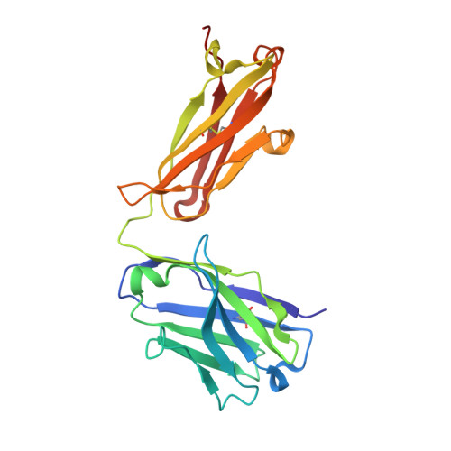
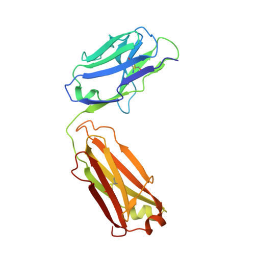
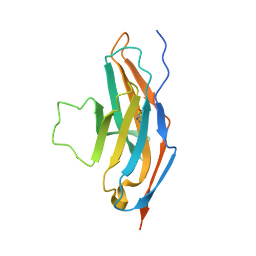
Identification of a monoclonal antibody that targets PD-1 in a manner requiring PD-1 Asn58 glycosylation.

Wang, M.Wang, J.Wang, R.Jiao, S.Wang, S.Zhang, J.Zhang, M.

(2019) Commun Biol 2: 392-392

  • DOI: https://doi.org/10.1038/s42003-019-0642-9
  • Primary Citation Related Structures: 
    6JJP

  • PubMed Abstract: 

    Programmed cell death 1 (PD-1) is inhibitory receptor and immune checkpoint protein. Blocking the interaction of PD-1 and its ligands PD-L1/ L2 is able to active T-cell-mediated antitumor response. Monoclonal antibody-based drugs targeting PD-1 pathway have exhibited great promise in cancer therapy. Here we show that MW11-h317, an anti-PD-1 monoclonal antibody, displays high affinity for PD-1 and blocks PD-1 interactions with PD-L1/L2. MW11-h317 can effectively induce T-cell-mediated immune response and inhibit tumor growth in mouse model. Crystal structure of PD-1/MW11-h317 Fab complex reveals that both the loops and glycosylation of PD-1 are involved in recognition and binding, in which Asn58 glycosylation plays a critical role. The unique glycan epitope in PD-1 to MW11-h317 is different from the first two approved clinical PD-1 antibodies, nivolumab and pembrolizumab. These results suggest MW11-h317 as a therapeutic monoclonal antibody of PD-1 glycosylation-targeting which may become efficient alternative for cancer therapy.


  • Organizational Affiliation
    • 1School of Life Sciences, Anhui University, 230601 Hefei, Anhui China.

Macromolecule Content 

  • Total Structure Weight: 128.9 kDa 
  • Atom Count: 8,592 
  • Modeled Residue Count: 1,102 
  • Deposited Residue Count: 1,162 
  • Unique protein chains: 3

Macromolecules

Find similar proteins by:|  3D Structure
Entity ID: 1
MoleculeChains  Sequence LengthOrganismDetailsImage
Heavy chain of MW11-h317
A, D
220Homo sapiensMutation(s): 0 
Find similar proteins by:|  3D Structure
Entity ID: 2
MoleculeChains  Sequence LengthOrganismDetailsImage
light chain of MW11-h317
B, E
214Homo sapiensMutation(s): 0 
Find similar proteins by:|  3D Structure
Entity ID: 3
MoleculeChains  Sequence LengthOrganismDetailsImage
Programmed cell death protein 1
C, F
147Homo sapiensMutation(s): 0 
Gene Names: PDCD1PD1
UniProt & NIH Common Fund Data Resources
Find proteins for Q15116 (Homo sapiens)
Explore Q15116 
Go to UniProtKB:  Q15116
PHAROS:  Q15116
GTEx:  ENSG00000188389 
Entity Groups
Sequence Clusters30% Identity50% Identity70% Identity90% Identity95% Identity100% Identity
UniProt GroupQ15116
Glycosylation
Glycosylation Sites: 3Go to GlyGen: Q15116-1
Sequence Annotations
Expand
Reference Sequence

Oligosaccharides

Help  
Entity ID: 4
MoleculeChains Length2D Diagram GlycosylationD Interactions
alpha-D-mannopyranose-(1-3)-beta-D-mannopyranose-(1-4)-2-acetamido-2-deoxy-beta-D-glucopyranose-(1-4)-[alpha-L-fucopyranose-(1-6)]2-acetamido-2-deoxy-beta-D-glucopyranose
G, H
5N-Glycosylation
Glycosylation Resources
GlyTouCan: G42466VF
GlyCosmos: G42466VF
GlyGen: G42466VF

Experimental Data & Validation

Experimental Data

  • Method: X-RAY DIFFRACTION
  • Resolution: 2.90 Å
  • R-Value Free:  0.247 (Depositor), 0.248 (DCC) 
  • R-Value Work:  0.207 (Depositor), 0.212 (DCC) 
  • R-Value Observed: 0.209 (Depositor) 
Space Group: P 1 2 1
Unit Cell:
Length ( Å )Angle ( ˚ )
a = 102.61α = 90
b = 54.219β = 113.92
c = 126.082γ = 90
Software Package:
Software NamePurpose
HKL-2000data reduction
HKL-2000data scaling
PHENIXrefinement
PDB_EXTRACTdata extraction
PHASERphasing

Structure Validation

View Full Validation Report


Ligand Structure Quality Assessment 


Entry History 

Deposition Data

Revision History  (Full details and data files)

  • Version 1.0: 2019-10-30
    Type: Initial release
  • Version 1.1: 2019-11-06
    Changes: Data collection, Derived calculations
  • Version 1.2: 2019-11-13
    Changes: Data collection, Database references
  • Version 2.0: 2020-07-29
    Type: Remediation
    Reason: Carbohydrate remediation
    Changes: Advisory, Atomic model, Data collection, Derived calculations, Structure summary
  • Version 2.1: 2023-11-22
    Changes: Data collection, Database references, Refinement description, Structure summary
  • Version 2.2: 2024-11-06
    Changes: Structure summary