6JHN

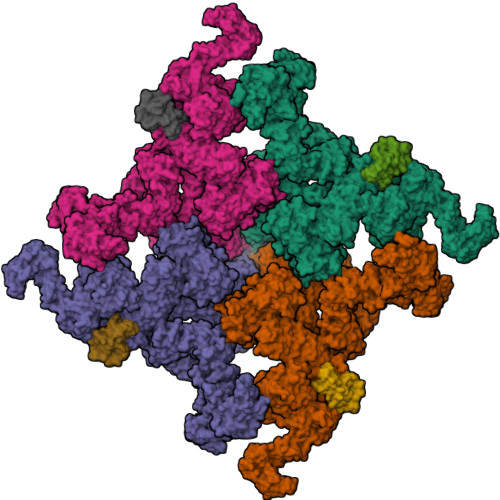
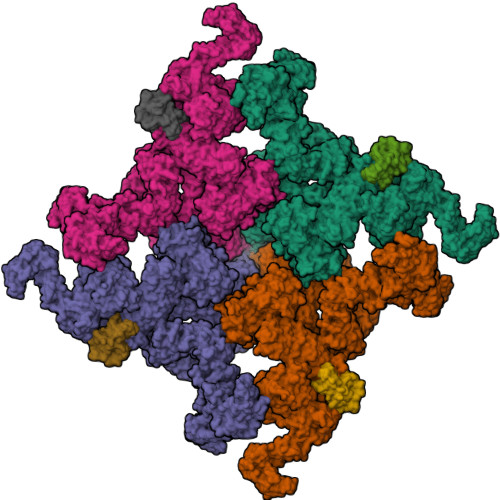
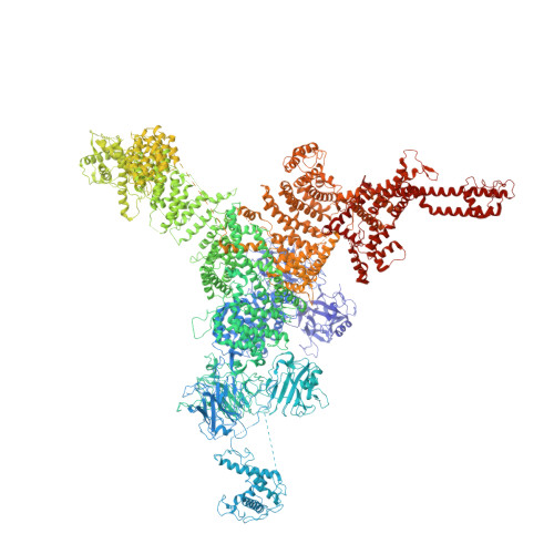
Structure of RyR2 (F/C/Ca2+ dataset)


Experimental Data Snapshot

  • Method: ELECTRON MICROSCOPY
  • Resolution: 4.50 Å
  • Aggregation State: PARTICLE 
  • Reconstruction Method: SINGLE PARTICLE 

wwPDB Validation   3D Report Full Report


This is version 1.2 of the entry. See complete history


Literature

Molecular basis for allosteric regulation of the type 2 ryanodine receptor channel gating by key modulators.

Chi, X.Gong, D.Ren, K.Zhou, G.Huang, G.Lei, J.Zhou, Q.Yan, N.

(2019) Proc Natl Acad Sci U S A 116: 25575-25582

  • DOI: https://doi.org/10.1073/pnas.1914451116
  • Primary Citation of Related Structures:  
    6JG3, 6JGZ, 6JH6, 6JHN

  • PubMed Abstract: 

    The type 2 ryanodine receptor (RyR2) is responsible for releasing Ca 2+ from the sarcoplasmic reticulum of cardiomyocytes, subsequently leading to muscle contraction. Here, we report 4 cryo-electron microscopy (cryo-EM) structures of porcine RyR2 bound to distinct modulators that, together with our published structures, provide mechanistic insight into RyR2 regulation. Ca 2+ alone induces a contraction of the central domain that facilitates the dilation of the S6 bundle but is insufficient to open the pore. The small-molecule agonist PCB95 helps Ca 2+ to overcome the barrier for opening. FKBP12.6 induces a relaxation of the central domain that decouples it from the S6 bundle, stabilizing RyR2 in a closed state even in the presence of Ca 2+ and PCB95. Although the channel is open when PCB95 is replaced by caffeine and adenosine 5'-triphosphate (ATP), neither of the modulators alone can sufficiently counter the antagonistic effect to open the channel. Our study marks an important step toward mechanistic understanding of the sophisticated regulation of this key channel whose aberrant activity engenders life-threatening cardiac disorders.


  • Organizational Affiliation

    Key Laboratory of Structural Biology of Zhejiang Province, School of Life Sciences, Westlake University, Hangzhou 310024, China.


Macromolecules
Find similar proteins by:  (by identity cutoff)  |  3D Structure
Entity ID: 1
MoleculeChains Sequence LengthOrganismDetailsImage
RyR2
A, C, E, G
4,968Sus scrofaMutation(s): 0 
Entity Groups  
Sequence Clusters30% Identity50% Identity70% Identity90% Identity95% Identity100% Identity
Sequence Annotations
Expand
  • Reference Sequence
Find similar proteins by:  (by identity cutoff)  |  3D Structure
Entity ID: 2
MoleculeChains Sequence LengthOrganismDetailsImage
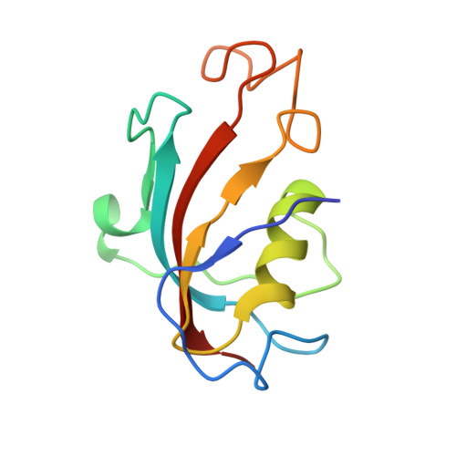
Peptidyl-prolyl cis-trans isomerase FKBP1B
B, D, F, H
108Homo sapiensMutation(s): 0 
Gene Names: FKBP1BFKBP12.6FKBP1LFKBP9OTK4
EC: 5.2.1.8
UniProt & NIH Common Fund Data Resources
Find proteins for P68106 (Homo sapiens)
Explore P68106 
Go to UniProtKB:  P68106
PHAROS:  P68106
GTEx:  ENSG00000119782 
Entity Groups  
Sequence Clusters30% Identity50% Identity70% Identity90% Identity95% Identity100% Identity
UniProt GroupP68106
Sequence Annotations
Expand
  • Reference Sequence
Small Molecules
Ligands 3 Unique
IDChains Name / Formula / InChI Key2D Diagram3D Interactions
CFF
Query on CFF

Download Ideal Coordinates CCD File 
K [auth A],
N [auth C],
Q [auth E],
T [auth G]
CAFFEINE
C8 H10 N4 O2
RYYVLZVUVIJVGH-UHFFFAOYSA-N
ZN
Query on ZN

Download Ideal Coordinates CCD File 
I [auth A],
L [auth C],
O [auth E],
R [auth G]
ZINC ION
Zn
PTFCDOFLOPIGGS-UHFFFAOYSA-N
CA
Query on CA

Download Ideal Coordinates CCD File 
J [auth A],
M [auth C],
P [auth E],
S [auth G]
CALCIUM ION
Ca
BHPQYMZQTOCNFJ-UHFFFAOYSA-N
Experimental Data & Validation

Experimental Data

  • Method: ELECTRON MICROSCOPY
  • Resolution: 4.50 Å
  • Aggregation State: PARTICLE 
  • Reconstruction Method: SINGLE PARTICLE 
EM Software:
TaskSoftware PackageVersion
RECONSTRUCTIONRELION2.0

Structure Validation

View Full Validation Report


Entry History 

Deposition Data

Revision History  (Full details and data files)

  • Version 1.0: 2019-12-11
    Type: Initial release
  • Version 1.1: 2020-01-01
    Changes: Database references
  • Version 1.2: 2024-03-27
    Changes: Data collection, Database references