6JD8 | pdb_00006jd8

Structure of a proline specific mutant of human cathepsin L


Experimental Data Snapshot

  • Method: X-RAY DIFFRACTION
  • Resolution: 1.46 Å
  • R-Value Free: 
    0.237 (Depositor), 0.248 (DCC) 
  • R-Value Work: 
    0.192 (Depositor) 
  • R-Value Observed: 
    0.193 (Depositor) 

Starting Model: experimental
View more details

wwPDB Validation 3D Report Full Report

Validation slider image for 6JD8

This is version 1.3 of the entry. See complete history

Literature

Structure-guided protein engineering of human cathepsin L for efficient collagenolytic activity.

Choudhury, D.Biswas, S.

(2021) Protein Eng Des Sel 34

  • DOI: https://doi.org/10.1093/protein/gzab005
  • Primary Citation Related Structures: 
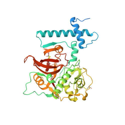
    6JD0, 6JD8

  • PubMed Abstract: 

    Engineering precise substrate specificity of proteases advances the potential to use them in biotechnological and therapeutic applications. Collagen degradation, a physiological process mediated by collagenases, is an integral part of extracellular matrix remodeling and when uncontrolled, implicated in different pathological conditions. Lysosomal cathepsin-K cleaves triple helical collagen fiber, whereas cathepsin-L cannot do so. In this study, we have imparted collagenolytic property to cathepsin-L, by systematically engineering proline-specificity and glycosaminoglycans (GAG)-binding surface in the protease. The proline-specific mutant shows high specificity for prolyl-peptidic substrate but is incapable of cleaving collagen. Engineering a GAG-binding surface on the proline-specific mutant enabled it to degrade type-I collagen in the presence of chondroitin-4-sulfate (C4-S). We also present the crystal structures of proline-specific (1.4 Å) and collagen-specific (1.8 Å) mutants. Finally docking studies with prolyl-peptidic substrate (Ala-Gly-Pro-Arg-Ala) at the active site and a C4-S molecule at the GAG-binding site enable us to identify key structural features responsible for collagenolytic activity of cysteine cathepsins.


  • Organizational Affiliation
    • Crystallography and Molecular Biology Division, Saha Institute of Nuclear Physics, 1/AF Bidhannagar, Kolkata 700 064, India.

Macromolecule Content 

  • Total Structure Weight: 41.88 kDa 
  • Atom Count: 2,878 
  • Modeled Residue Count: 314 
  • Deposited Residue Count: 360 
  • Unique protein chains: 1

Macromolecules

Find similar proteins by:|  3D Structure
Entity ID: 1
MoleculeChains  Sequence LengthOrganismDetailsImage
Cathepsin L1360Homo sapiensMutation(s): 5 
Gene Names: CTSLCTSL1
EC: 3.4.22.15
UniProt & NIH Common Fund Data Resources
Find proteins for P07711 (Homo sapiens)
Explore P07711 
Go to UniProtKB:  P07711
PHAROS:  P07711
GTEx:  ENSG00000135047 
Entity Groups
Sequence Clusters30% Identity50% Identity70% Identity90% Identity95% Identity100% Identity
UniProt GroupP07711
Sequence Annotations
Expand
Reference Sequence

Small Molecules

Ligands 5 Unique
IDChains Name / Formula / InChI Key2D Diagram3D Interactions
PGE

Query on PGE


Download:Ideal Coordinates CCD File
Q [auth A]TRIETHYLENE GLYCOL
C6 H14 O4
ZIBGPFATKBEMQZ-UHFFFAOYSA-N
PEG

Query on PEG


Download:Ideal Coordinates CCD File
O [auth A]DI(HYDROXYETHYL)ETHER
C4 H10 O3
MTHSVFCYNBDYFN-UHFFFAOYSA-N
GOL

Query on GOL


Download:Ideal Coordinates CCD File
B [auth A]GLYCEROL
C3 H8 O3
PEDCQBHIVMGVHV-UHFFFAOYSA-N
EDO

Query on EDO


Download:Ideal Coordinates CCD File
P [auth A]1,2-ETHANEDIOL
C2 H6 O2
LYCAIKOWRPUZTN-UHFFFAOYSA-N
EOH

Query on EOH


Download:Ideal Coordinates CCD File
C [auth A]
D [auth A]
E [auth A]
F [auth A]
G [auth A]
C [auth A],
D [auth A],
E [auth A],
F [auth A],
G [auth A],
H [auth A],
I [auth A],
J [auth A],
K [auth A],
L [auth A],
M [auth A],
N [auth A]
ETHANOL
C2 H6 O
LFQSCWFLJHTTHZ-UHFFFAOYSA-N

Experimental Data & Validation

Experimental Data

  • Method: X-RAY DIFFRACTION
  • Resolution: 1.46 Å
  • R-Value Free:  0.237 (Depositor), 0.248 (DCC) 
  • R-Value Work:  0.192 (Depositor) 
  • R-Value Observed: 0.193 (Depositor) 
Space Group: P 21 21 21
Unit Cell:
Length ( Å )Angle ( ˚ )
a = 42.22α = 90
b = 67.584β = 90
c = 103.239γ = 90
Software Package:
Software NamePurpose
HKL-2000data reduction
HKL-2000data scaling
PHENIXrefinement
PDB_EXTRACTdata extraction
PHASERphasing

Structure Validation

View Full Validation Report


Entry History 

& Funding Information

Deposition Data


Funding OrganizationLocationGrant Number
Department of Science & Technology (India)India--

Revision History  (Full details and data files)

  • Version 1.0: 2020-02-05
    Type: Initial release
  • Version 1.1: 2022-10-19
    Changes: Database references
  • Version 1.2: 2023-11-29
    Changes: Data collection, Refinement description
  • Version 1.3: 2024-11-13
    Changes: Structure summary