6JBE | pdb_00006jbe

Crystal structure of ABC transporter alpha-glycoside-binding mutant protein W287A in complex with glucose


Experimental Data Snapshot

  • Method: X-RAY DIFFRACTION
  • Resolution: 1.75 Å
  • R-Value Free: 
    0.176 (Depositor), 0.182 (DCC) 
  • R-Value Work: 
    0.139 (Depositor), 0.154 (DCC) 
  • R-Value Observed: 
    0.141 (Depositor) 

Starting Model: experimental
View more details

wwPDB Validation 3D Report Full Report

Validation slider image for 6JBE

Ligand Structure Quality Assessment 


This is version 1.3 of the entry. See complete history

Literature

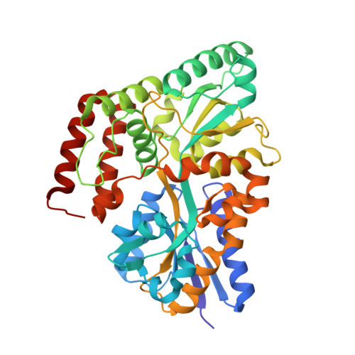
Structural and thermodynamic correlation illuminates the selective transport mechanism of disaccharide alpha-glycosides through ABC transporter.

Chandravanshi, M.Gogoi, P.Kanaujia, S.P.

(2020) FEBS J 287: 1576-1597

  • DOI: https://doi.org/10.1111/febs.15093
  • Primary Citation Related Structures: 
    6J9W, 6J9Y, 6JAD, 6JAG, 6JAH, 6JAI, 6JAL, 6JAM, 6JAN, 6JAO, 6JAP, 6JAQ, 6JAR, 6JAZ, 6JB0, 6JB4, 6JBA, 6JBB, 6JBE

  • PubMed Abstract: 

    Carbohydrate (or sugar) molecules are extremely diverse regarding their length, linkage and epimeric state. Selective acquisition of these molecules inside the cell is achieved by the substrate (or solute)-binding protein of ATP-binding cassette (ABC) transport system. However, the molecular mechanism underlying the selective transport of diverse carbohydrates remains unclear mainly owing to their structural complexity and stereochemistry. This study reports crystal structures of an α-glycoside-binding protein (αGlyBP, ORF ID: TTHA0356 from Thermus thermophilus HB8) in complex with disaccharide α-glycosides namely trehalose (α-1,1), sucrose (α-1,2), maltose (α-1,4), palatinose (α-1,6) and glucose within a resolution range of 1.6-2.0 Å. Despite transporting multiple types of sugars, αGlyBP maintains its stereoselectivity for both glycosidic linkage as well as an epimeric hydroxyl group. Out of the two subsites identified in the active-site pocket, subsite B which accommodates the glucose and glycosyl unit of disaccharide α-glycosides is highly conserved. In addition, structural data confirms the paradoxical behavior of glucose, where it replaces the high-affinity ligand(s) (disaccharide α-glycosides) from the active site of the protein. Comparative assessment of open and closed conformations of αGlyBP along with mutagenic and thermodynamic studies identifies the hinge region as the first interaction site for the ligands. On the other hand, encapsulation of ligand inside the active site is achieved through the N-terminal domain (NTD) movement, whereas the C-terminal domain (CTD) of αGlyBP is identified to be rigid and postulated to be responsible for maintaining the interaction with the transmembrane domain (TMD) during substrate translocation. DATABASE: Structural data are available in RCSB Protein Data Bank under the accession number(s) 6J9W, 6J9Y, 6JAD, 6JAG, 6JAH, 6JAI, 6JAL, 6JAM, 6JAN, 6JAO, 6JAP, 6JAQ, 6JAR, 6JAZ, 6JB0, 6JB4, 6JBA, 6JBB and 6JBE.


  • Organizational Affiliation
    • Department of Biosciences and Bioengineering, Indian Institute of Technology Guwahati, India.

Macromolecule Content 

  • Total Structure Weight: 46.82 kDa 
  • Atom Count: 3,870 
  • Modeled Residue Count: 404 
  • Deposited Residue Count: 408 
  • Unique protein chains: 1

Macromolecules

Find similar proteins by:|  3D Structure
Entity ID: 1
MoleculeChains  Sequence LengthOrganismDetailsImage
ABC transporter, periplasmic substrate-binding protein408Thermus thermophilus HB8Mutation(s): 1 
Gene Names: TTHA0356
UniProt
Find proteins for Q5SLD7 (Thermus thermophilus (strain ATCC 27634 / DSM 579 / HB8))
Explore Q5SLD7 
Go to UniProtKB:  Q5SLD7
Entity Groups
Sequence Clusters30% Identity50% Identity70% Identity90% Identity95% Identity100% Identity
UniProt GroupQ5SLD7
Sequence Annotations
Expand
Reference Sequence

Small Molecules

Ligands 6 Unique
IDChains Name / Formula / InChI Key2D Diagram3D Interactions
CIT

Query on CIT


Download:Ideal Coordinates CCD File
C [auth A]CITRIC ACID
C6 H8 O7
KRKNYBCHXYNGOX-UHFFFAOYSA-N
GLC
(Subject of Investigation/LOI)

Query on GLC


Download:Ideal Coordinates CCD File
B [auth A]alpha-D-glucopyranose
C6 H12 O6
WQZGKKKJIJFFOK-DVKNGEFBSA-N
PEG

Query on PEG


Download:Ideal Coordinates CCD File
F [auth A]DI(HYDROXYETHYL)ETHER
C4 H10 O3
MTHSVFCYNBDYFN-UHFFFAOYSA-N
GOL

Query on GOL


Download:Ideal Coordinates CCD File
D [auth A],
E [auth A]
GLYCEROL
C3 H8 O3
PEDCQBHIVMGVHV-UHFFFAOYSA-N
EDO

Query on EDO


Download:Ideal Coordinates CCD File
G [auth A]1,2-ETHANEDIOL
C2 H6 O2
LYCAIKOWRPUZTN-UHFFFAOYSA-N
CO2

Query on CO2


Download:Ideal Coordinates CCD File
H [auth A]CARBON DIOXIDE
C O2
CURLTUGMZLYLDI-UHFFFAOYSA-N

Experimental Data & Validation

Experimental Data

  • Method: X-RAY DIFFRACTION
  • Resolution: 1.75 Å
  • R-Value Free:  0.176 (Depositor), 0.182 (DCC) 
  • R-Value Work:  0.139 (Depositor), 0.154 (DCC) 
  • R-Value Observed: 0.141 (Depositor) 
Space Group: P 41 21 2
Unit Cell:
Length ( Å )Angle ( ˚ )
a = 84.99α = 90
b = 84.99β = 90
c = 145.83γ = 90
Software Package:
Software NamePurpose
HKL-3000data collection
iMOSFLMdata reduction
Aimlessdata scaling
PHASERphasing
REFMACrefinement
Cootmodel building
PDB_EXTRACTdata extraction

Structure Validation

View Full Validation Report


Ligand Structure Quality Assessment 


Entry History 

& Funding Information

Deposition Data


Funding OrganizationLocationGrant Number
Department of Biotechnology (India)IndiaBT/563/NE/U-Excel/2016

Revision History  (Full details and data files)

  • Version 1.0: 2019-10-30
    Type: Initial release
  • Version 1.1: 2020-05-06
    Changes: Data collection, Database references
  • Version 1.2: 2020-07-29
    Type: Remediation
    Reason: Carbohydrate remediation
    Changes: Data collection, Derived calculations, Structure summary
  • Version 1.3: 2023-11-22
    Changes: Data collection, Database references, Refinement description, Structure summary