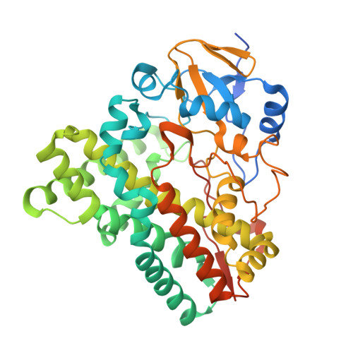
6J85

Crystal structure of HinD apo


Experimental Data Snapshot

  • Method: X-RAY DIFFRACTION
  • Resolution: 2.20 Å
  • R-Value Free: 0.226 
  • R-Value Work: 0.170 
  • R-Value Observed: 0.182 

Starting Model: experimental
View more details

wwPDB Validation   3D Report Full Report


Ligand Structure Quality Assessment 


This is version 1.2 of the entry. See complete history


Literature

Molecular basis for the P450-catalyzed C-N bond formation in indolactam biosynthesis.

He, F.Mori, T.Morita, I.Nakamura, H.Alblova, M.Hoshino, S.Awakawa, T.Abe, I.

(2019) Nat Chem Biol 15: 1206-1213

  • DOI: https://doi.org/10.1038/s41589-019-0380-9
  • Primary Citation of Related Structures:  
    6J82, 6J83, 6J84, 6J85, 6J86, 6J87, 6J88

  • PubMed Abstract: 

    The catalytic versatility of cytochrome P450 monooxygenases is remarkable. Here, we present mechanistic and structural characterizations of TleB from Streptomyces blastmyceticus and its homolog HinD from Streptoalloteichus hindustanus, which catalyze unusual intramolecular C-N bond formation to generate indolactam V from the dipeptide N-methylvalyl-tryptophanol. In vitro analyses demonstrated that both P450s exhibit promiscuous substrate specificity, and modification of the N13-methyl group resulted in the formation of indole-fused 6/5/6 tricyclic products. Furthermore, X-ray crystal structures in complex with substrates and structure-based mutagenesis revealed the intimate structural details of the enzyme reactions. We propose that the generation of a diradical species is critical for the indolactam formation, and that the intramolecular C(sp 2 )-H amination is initiated by the abstraction of the N1 indole hydrogen. After indole radical repositioning and subsequent removal of the N13 hydrogen, the coupling of the properly-folded diradical leads to the formation of the C4-N13 bond of indolactam.


  • Organizational Affiliation

    Graduate School of Pharmaceutical Sciences, The University of Tokyo, Tokyo, Japan.


Macromolecules
Find similar proteins by:  (by identity cutoff)  |  3D Structure
Entity ID: 1
MoleculeChains Sequence LengthOrganismDetailsImage
Nocardicin N-oxygenase
A, B
435Streptoalloteichus hindustanusMutation(s): 0 
Gene Names: SAMN05444320_102263
UniProt
Find proteins for A0A1M4Y7D5 (Streptoalloteichus hindustanus)
Explore A0A1M4Y7D5 
Go to UniProtKB:  A0A1M4Y7D5
Entity Groups  
Sequence Clusters30% Identity50% Identity70% Identity90% Identity95% Identity100% Identity
UniProt GroupA0A1M4Y7D5
Sequence Annotations
Expand
  • Reference Sequence
Experimental Data & Validation

Experimental Data

  • Method: X-RAY DIFFRACTION
  • Resolution: 2.20 Å
  • R-Value Free: 0.226 
  • R-Value Work: 0.170 
  • R-Value Observed: 0.182 
  • Space Group: P 1 21 1
Unit Cell:
Length ( Å )Angle ( ˚ )
a = 49.356α = 90
b = 152.647β = 115.85
c = 56.394γ = 90
Software Package:
Software NamePurpose
PHENIXrefinement
XDSdata reduction
Aimlessdata scaling
PHASERphasing

Structure Validation

View Full Validation Report


Ligand Structure Quality Assessment 


Entry History 

Deposition Data

Revision History  (Full details and data files)

  • Version 1.0: 2019-08-07
    Type: Initial release
  • Version 1.1: 2021-02-17
    Changes: Database references, Derived calculations
  • Version 1.2: 2023-11-22
    Changes: Data collection, Database references, Refinement description