6J22 | pdb_00006j22

Crystal structure of Bi-functional enzyme


Experimental Data Snapshot

  • Method: X-RAY DIFFRACTION
  • Resolution: 2.20 Å
  • R-Value Free: 
    0.258 (Depositor), 0.268 (DCC) 
  • R-Value Work: 
    0.196 (Depositor) 
  • R-Value Observed: 
    0.199 (Depositor) 

wwPDB Validation 3D Report Full Report

Validation slider image for 6J22

This is version 1.1 of the entry. See complete history

Literature

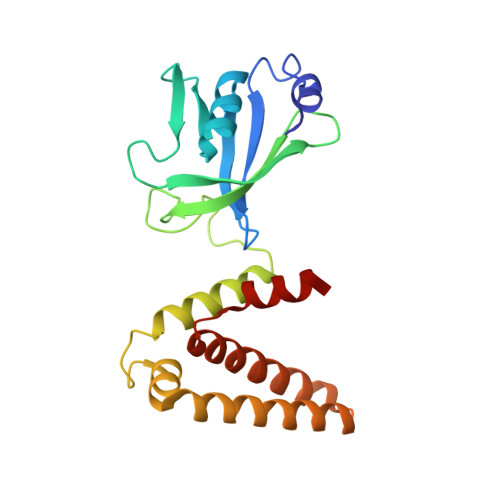
Structural analysis of Shigella flexneri bi-functional enzyme HisIE in histidine biosynthesis.

Wang, Y.Zhang, F.Nie, Y.Shang, G.Zhang, H.

(2019) Biochem Biophys Res Commun 516: 540-545

  • DOI: https://doi.org/10.1016/j.bbrc.2019.06.099
  • Primary Citation Related Structures: 
    6J22, 6J2L

  • PubMed Abstract: 

    Histidine biosynthesis, which is absent in animals, was shown to be highly conserved among gram-negative bacteria, thus making it an attractive target for antibiotic design. There are many fusion forms of enzymes in the histidine biosynthetic pathway and people still have limited knowledge about their domain organizations and catalytic mechanisms, due to the lack of structural information. Here we report the first crystal structure of Shigella flexneri bi-functional enzyme HisIE (SfHisIE) that functions in the 2nd and 3rd steps in the histidine biosynthetic pathway. This structure shows that HisIE exists as dimers with two loops (fusion loop) connecting the individual dimer of HisE and HisI in its N-terminus and C-terminus respectively. Our mutagenesis study shows mutations in this fusion loop are lethal for bacteria indicating the advantage of gene fusion in Histidine biosynthesis. Structural analysis revealed several highly conserved residues in the putative ligand binding grooves of HisE and HisI, showing an evolutionarily conserved catalytic mechanism shared among gram negative-bacteria.


  • Organizational Affiliation
    • Shanghai Institute for Advanced Immunochemical Studies (SIAIS), ShanghaiTech University, Shanghai, China.

Macromolecule Content 

  • Total Structure Weight: 45.62 kDa 
  • Atom Count: 3,247 
  • Modeled Residue Count: 385 
  • Deposited Residue Count: 404 
  • Unique protein chains: 1

Macromolecules

Find similar proteins by:|  3D Structure
Entity ID: 1
MoleculeChains  Sequence LengthOrganismDetailsImage
Histidine biosynthesis bifunctional protein HisIE
A, B
202Shigella flexneriMutation(s): 0 
Gene Names: hisIhisIESF2088S2209
EC: 3.5.4.19 (PDB Primary Data), 3.6.1.31 (PDB Primary Data)
UniProt
Find proteins for P37793 (Shigella flexneri)
Explore P37793 
Go to UniProtKB:  P37793
Entity Groups
Sequence Clusters30% Identity50% Identity70% Identity90% Identity95% Identity100% Identity
UniProt GroupP37793
Sequence Annotations
Expand
Reference Sequence

Experimental Data & Validation

Experimental Data

  • Method: X-RAY DIFFRACTION
  • Resolution: 2.20 Å
  • R-Value Free:  0.258 (Depositor), 0.268 (DCC) 
  • R-Value Work:  0.196 (Depositor) 
  • R-Value Observed: 0.199 (Depositor) 
Space Group: C 1 2 1
Unit Cell:
Length ( Å )Angle ( ˚ )
a = 168.711α = 90
b = 45.195β = 112.46
c = 65.8γ = 90
Software Package:
Software NamePurpose
REFMACrefinement
HKL-2000data reduction
HKL-2000data scaling
PHASERphasing

Structure Validation

View Full Validation Report


Entry History 

Deposition Data

Revision History  (Full details and data files)

  • Version 1.0: 2020-01-01
    Type: Initial release
  • Version 1.1: 2024-03-27
    Changes: Data collection, Database references