6IVE | pdb_00006ive

Molecular structure of a thermostable and a Zinc ion binding gamma-class carbonic anhydrase


Experimental Data Snapshot

  • Method: X-RAY DIFFRACTION
  • Resolution: 2.30 Å
  • R-Value Free: 
    0.223 (Depositor), 0.223 (DCC) 
  • R-Value Work: 
    0.181 (Depositor), 0.182 (DCC) 
  • R-Value Observed: 
    0.183 (Depositor) 

Starting Model: experimental
View more details

wwPDB Validation 3D Report Full Report

Validation slider image for 6IVE

This is version 1.2 of the entry. See complete history

Literature

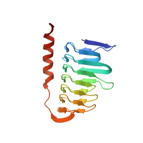
Molecular structure of thermostable and zinc-ion-binding gamma-class carbonic anhydrases.

Wang, W.Zhang, Y.Wang, L.Jing, Q.Wang, X.Xi, X.Zhao, X.Wang, H.

(2019) Biometals 32: 317-328

  • DOI: https://doi.org/10.1007/s10534-019-00190-8
  • Primary Citation Related Structures: 
    6IVE

  • PubMed Abstract: 

    The γ-class carbonic anhydrases (γ-CAs) mainly come from methanogens methane-producing bacteria that grow in hot springs and catalyze the interconversion of carbon dioxide and water to bicarbonate and protons. Here, the γ-CA from Thermus thermophilus HB8 (γ-TtCA) was expressed and purified, its crystal structure was determined at 2.3 Å resolution in space group P1. The asymmetric unit contains two trimers and six catalytic Zn 2+ . In general, the fold of the protein is similar to those of homologous enzymes from Geobacillus Kaustophilus, Bacillus Cereus, Methanosarcina Thermophila and others. Each monomer comprises a triangular prism-like structure consisting of a left-handed β-helix and a C-terminal α-helix. The catalytic Zn 2+ bound to three histidines and a phosphate radical in a tetrahedral fashion. It is located at the interface between the two monomers. Inductively coupled plasma mass spectrometry measurements further suggest that the molar ratio of zinc ions and protein molecules is 1:1. The structure revealed a novel different region situated between the left-handed β-helix and the C-terminal α-helix. Compared to previously reported structures, half of the C-terminal α-helix was replaced with a long loop in this structure. The purified γ-TtCA exhibits no significant carbonic anhydrase activity compared to α-class carbonic anhydrases. This study provides insight into the structural diversity of γ-CAs with potential function for γ-CAs.


  • Organizational Affiliation
    • Key Laboratory of Chemical Biology and Molecular Engineering of Education Ministry, Institute of Molecular Science, Shanxi University, Taiyuan, 030006, China. wangwm@sxu.edu.cn.

Macromolecule Content 

  • Total Structure Weight: 105.83 kDa 
  • Atom Count: 7,678 
  • Modeled Residue Count: 1,003 
  • Deposited Residue Count: 1,026 
  • Unique protein chains: 1

Macromolecules

Find similar proteins by:|  3D Structure
Entity ID: 1
MoleculeChains  Sequence LengthOrganismDetailsImage
Ferripyochelin-binding protein
A, B, C, D, E
A, B, C, D, E, F
171Thermus thermophilus HB8Mutation(s): 0 
Gene Names: TTHA1879
UniProt
Find proteins for Q5SH51 (Thermus thermophilus (strain ATCC 27634 / DSM 579 / HB8))
Explore Q5SH51 
Go to UniProtKB:  Q5SH51
Entity Groups
Sequence Clusters30% Identity50% Identity70% Identity90% Identity95% Identity100% Identity
UniProt GroupQ5SH51
Sequence Annotations
Expand
Reference Sequence

Small Molecules

Ligands 2 Unique
IDChains Name / Formula / InChI Key2D Diagram3D Interactions
PO4

Query on PO4


Download:Ideal Coordinates CCD File
H [auth A]
I [auth A]
L [auth C]
N [auth D]
O [auth D]
H [auth A],
I [auth A],
L [auth C],
N [auth D],
O [auth D],
Q [auth E]
PHOSPHATE ION
O4 P
NBIIXXVUZAFLBC-UHFFFAOYSA-K
ZN

Query on ZN


Download:Ideal Coordinates CCD File
G [auth A]
J [auth B]
K [auth C]
M [auth D]
P [auth E]
G [auth A],
J [auth B],
K [auth C],
M [auth D],
P [auth E],
R [auth F]
ZINC ION
Zn
PTFCDOFLOPIGGS-UHFFFAOYSA-N

Experimental Data & Validation

Experimental Data

  • Method: X-RAY DIFFRACTION
  • Resolution: 2.30 Å
  • R-Value Free:  0.223 (Depositor), 0.223 (DCC) 
  • R-Value Work:  0.181 (Depositor), 0.182 (DCC) 
  • R-Value Observed: 0.183 (Depositor) 
Space Group: P 1
Unit Cell:
Length ( Å )Angle ( ˚ )
a = 48.687α = 75.22
b = 69.232β = 74.39
c = 83.334γ = 89.2
Software Package:
Software NamePurpose
PHENIXrefinement
HKL-3000data reduction
HKL-3000data scaling
PHENIXphasing

Structure Validation

View Full Validation Report


Entry History 

& Funding Information

Deposition Data


Funding OrganizationLocationGrant Number
National Natural Science Foundation of ChinaChina21601112, 21671125

Revision History  (Full details and data files)

  • Version 1.0: 2019-04-03
    Type: Initial release
  • Version 1.1: 2019-04-10
    Changes: Data collection, Database references
  • Version 1.2: 2023-11-22
    Changes: Data collection, Database references, Derived calculations, Refinement description