6IUV

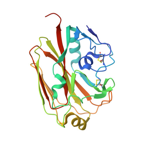
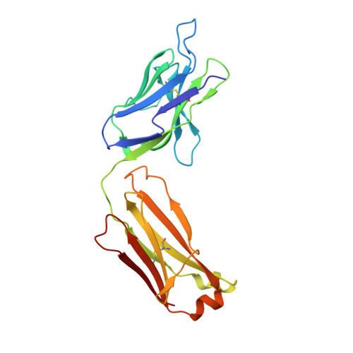
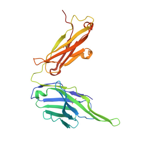
Crystal structure of influenza A virus H5 hemagglutinin globular head in complex with the Fab of antibody 3C11


Experimental Data Snapshot

  • Method: X-RAY DIFFRACTION
  • Resolution: 2.33 Å
  • R-Value Free: 0.246 
  • R-Value Work: 0.207 
  • R-Value Observed: 0.209 

Starting Model: experimental
View more details

wwPDB Validation   3D Report Full Report


This is version 2.1 of the entry. See complete history


Literature

Structural and functional definition of a vulnerable site on the hemagglutinin of highly pathogenic avian influenza A virus H5N1.

Wang, P.Zuo, Y.Sun, J.Zuo, T.Zhang, S.Guo, S.Shi, X.Liang, M.Zhou, P.Zhang, L.Wang, X.

(2019) J Biol Chem 294: 4290-4303

  • DOI: https://doi.org/10.1074/jbc.RA118.007008
  • Primary Citation of Related Structures:  
    6IUT, 6IUV

  • PubMed Abstract: 

    Most neutralizing antibodies against highly pathogenic avian influenza A virus H5N1 recognize the receptor-binding site (RBS) on the globular head domain and the stem of H5N1 hemagglutinin (HA). Through comprehensive analysis of multiple human protective antibodies, we previously identified four vulnerable sites (VS1-VS4) on the globular head domain. Among them, the VS1, occupying the opposite side of the RBS on the same HA, was defined by the epitope of antibody 65C6. In this study, we report the crystal structures of two additional human H5N1 antibodies isolated from H5N1-infected individuals, 3C11 and AVFluIgG01, bound to the head at 2.33- and 2.30-Å resolution, respectively. These two new antibody epitopes have large overlap with and extend beyond the original VS1. Site-directed mutagenesis experiments identified eight pivotal residues (Ser-126b, Lys-165, Arg-166, Ser-167, Tyr-168, Asn-169, Thr-171, and Asn-172) critical for 65C6-, 3C11-, and AVFluIgG01-binding and neutralization activities. These residues formed a unique "Y"-shaped surface on H5N1 globular head and are highly conserved among H5N1 viruses. Our results further support the existence of a vulnerable site distinct from the RBS and the stem region of H5N1 HA, and future design of immunogens should take this particular site into consideration.


  • Organizational Affiliation

    From The Ministry of Education Key Laboratory of Protein Science, Beijing Advanced Innovation Center for Structural Biology, Collaborative Innovation Center for Biotherapy, School of Life Sciences, Tsinghua University, Beijing 100084, China.


Macromolecules
Find similar proteins by:  (by identity cutoff)  |  3D Structure
Entity ID: 1
MoleculeChains Sequence LengthOrganismDetailsImage
HemagglutininA,
D [auth B]
224Influenza A virus (A/Hong Kong/482/97(H5N1))Mutation(s): 0 
Gene Names: H5HA
UniProt
Find proteins for Q77XR4 (Influenza A virus)
Explore Q77XR4 
Go to UniProtKB:  Q77XR4
Entity Groups  
Sequence Clusters30% Identity50% Identity70% Identity90% Identity95% Identity100% Identity
UniProt GroupQ77XR4
Glycosylation
Glycosylation Sites: 1
Sequence Annotations
Expand
  • Reference Sequence
Find similar proteins by:  (by identity cutoff)  |  3D Structure
Entity ID: 2
MoleculeChains Sequence LengthOrganismDetailsImage
3C11 Light ChainB [auth L],
E [auth D]
218Homo sapiensMutation(s): 0 
Entity Groups  
Sequence Clusters30% Identity50% Identity70% Identity90% Identity95% Identity100% Identity
Sequence Annotations
Expand
  • Reference Sequence
Find similar proteins by:  (by identity cutoff)  |  3D Structure
Entity ID: 3
MoleculeChains Sequence LengthOrganismDetailsImage
3C11 Heavy ChainC [auth H],
F [auth C]
229Homo sapiensMutation(s): 0 
Entity Groups  
Sequence Clusters30% Identity50% Identity70% Identity90% Identity95% Identity100% Identity
Sequence Annotations
Expand
  • Reference Sequence
Oligosaccharides

Help

Entity ID: 4
MoleculeChains Length2D Diagram Glycosylation3D Interactions
2-acetamido-2-deoxy-beta-D-glucopyranose-(1-4)-[alpha-L-fucopyranose-(1-6)]2-acetamido-2-deoxy-beta-D-glucopyranoseG [auth E],
H [auth F]
3N-Glycosylation
Glycosylation Resources
GlyTouCan:  G21290RB
GlyCosmos:  G21290RB
GlyGen:  G21290RB
Experimental Data & Validation

Experimental Data

  • Method: X-RAY DIFFRACTION
  • Resolution: 2.33 Å
  • R-Value Free: 0.246 
  • R-Value Work: 0.207 
  • R-Value Observed: 0.209 
  • Space Group: P 1 21 1
Unit Cell:
Length ( Å )Angle ( ˚ )
a = 126.78α = 90
b = 62.236β = 117.21
c = 127.827γ = 90
Software Package:
Software NamePurpose
PHENIXrefinement
HKL-2000data reduction
HKL-2000data scaling
PHASERphasing

Structure Validation

View Full Validation Report


Entry History & Funding Information

Deposition Data


Funding OrganizationLocationGrant Number
National Natural Science Foundation of ChinaChina81530065
National Natural Science Foundation of ChinaChina81661128042
National Natural Science Foundation of ChinaChina31470751
National Natural Science Foundation of ChinaChinaU1405228
Ministry of Science and Technology (China)China2016YFD0500307
Ministry of Science and Technology (China)China2012ZX10001-004
Ministry of Science and Technology (China)China2012ZX10001-006
Ministry of Science and Technology (China)China2012ZX10001-009
Ministry of Science and Technology (China)China2017ZX10201-101
Ministry of Science and Technology (China)China2014CB542500-03

Revision History  (Full details and data files)

  • Version 1.0: 2019-01-16
    Type: Initial release
  • Version 1.1: 2019-02-20
    Changes: Data collection, Database references
  • Version 1.2: 2019-04-10
    Changes: Data collection, Database references
  • Version 2.0: 2020-07-29
    Type: Remediation
    Reason: Carbohydrate remediation
    Changes: Atomic model, Data collection, Derived calculations, Structure summary
  • Version 2.1: 2023-11-22
    Changes: Data collection, Database references, Derived calculations, Refinement description, Structure summary