6ITO | pdb_00006ito

Crystal structure of pyruvate kinase (PYK) from Mycobacterium tuberculosis in complex with Oxalate, AMP and inhibitor Ribose 5-Phosphate


Experimental Data Snapshot

  • Method: X-RAY DIFFRACTION
  • Resolution: 2.55 Å
  • R-Value Free: 
    0.248 (Depositor), 0.246 (DCC) 
  • R-Value Work: 
    0.206 (Depositor), 0.208 (DCC) 
  • R-Value Observed: 
    0.208 (Depositor) 

Starting Model: experimental
View more details

wwPDB Validation 3D Report Full Report

Validation slider image for 6ITO

Ligand Structure Quality Assessment 


This is version 1.3 of the entry. See complete history

Literature

Pyruvate Kinase Regulates the Pentose-Phosphate Pathway in Response to Hypoxia in Mycobacterium tuberculosis.

Zhong, W.Guo, J.Cui, L.Chionh, Y.H.Li, K.El Sahili, A.Cai, Q.Yuan, M.Michels, P.A.M.Fothergill-Gilmore, L.A.Walkinshaw, M.D.Mu, Y.Lescar, J.Dedon, P.C.

(2019) J Mol Biology 431: 3690-3705

  • DOI: https://doi.org/10.1016/j.jmb.2019.07.033
  • Primary Citation Related Structures: 
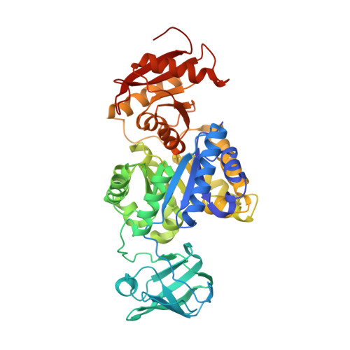
    6ITO

  • PubMed Abstract: 

    In response to the stress of infection, Mycobacterium tuberculosis (Mtb) reprograms its metabolism to accommodate nutrient and energetic demands in a changing environment. Pyruvate kinase (PYK) is an essential glycolytic enzyme in the phosphoenolpyruvate-pyruvate-oxaloacetate node that is a central switch point for carbon flux distribution. Here we show that the competitive binding of pentose monophosphate inhibitors or the activator glucose 6-phosphate (G6P) to MtbPYK tightly regulates the metabolic flux. Intriguingly, pentose monophosphates were found to share the same binding site with G6P. The determination of a crystal structure of MtbPYK with bound ribose 5-phosphate (R5P), combined with biochemical analyses and molecular dynamic simulations, revealed that the allosteric inhibitor pentose monophosphate increases PYK structural dynamics, weakens the structural network communication, and impairs substrate binding. G6P, on the other hand, primes and activates the tetramer by decreasing protein flexibility and strengthening allosteric coupling. Therefore, we propose that MtbPYK uses these differences in conformational dynamics to up- and down-regulate enzymic activity. Importantly, metabolome profiling in mycobacteria reveals a significant increase in the levels of pentose monophosphate during hypoxia, which provides insights into how PYK uses dynamics of the tetramer as a competitive allosteric mechanism to retard glycolysis and facilitate metabolic reprogramming toward the pentose-phosphate pathway for achieving redox balance and an anticipatory metabolic response in Mtb.


  • Organizational Affiliation
    • Antimicrobial Resistance Interdisciplinary Research Group, Singapore-MIT Alliance for Research and Technology, 1 CREATE Way, 138602, Singapore; NTU Institute of Structural Biology, Nanyang Technological University, 636921, Singapore.

Macromolecule Content 

  • Total Structure Weight: 206.82 kDa 
  • Atom Count: 14,921 
  • Modeled Residue Count: 1,884 
  • Deposited Residue Count: 1,900 
  • Unique protein chains: 1

Macromolecules

Find similar proteins by:|  3D Structure
Entity ID: 1
MoleculeChains  Sequence LengthOrganismDetailsImage
Pyruvate kinase
A, B, C, D
475Mycobacterium tuberculosis H37RvMutation(s): 0 
Gene Names: pykpykARv1617MTCY01B2.09
EC: 2.7.1.40
UniProt
Find proteins for P9WKE5 (Mycobacterium tuberculosis (strain ATCC 25618 / H37Rv))
Explore P9WKE5 
Go to UniProtKB:  P9WKE5
Entity Groups
Sequence Clusters30% Identity50% Identity70% Identity90% Identity95% Identity100% Identity
UniProt GroupP9WKE5
Sequence Annotations
Expand
Reference Sequence

Small Molecules

Ligands 4 Unique
IDChains Name / Formula / InChI Key2D Diagram3D Interactions
AMP

Query on AMP


Download:Ideal Coordinates CCD File
H [auth A],
L [auth B],
P [auth C],
T [auth D]
ADENOSINE MONOPHOSPHATE
C10 H14 N5 O7 P
UDMBCSSLTHHNCD-KQYNXXCUSA-N
R5P

Query on R5P


Download:Ideal Coordinates CCD File
G [auth A],
K [auth B],
O [auth C],
S [auth D]
RIBOSE-5-PHOSPHATE
C5 H11 O8 P
PPQRONHOSHZGFQ-LMVFSUKVSA-N
OXL

Query on OXL


Download:Ideal Coordinates CCD File
F [auth A],
J [auth B],
N [auth C],
R [auth D]
OXALATE ION
C2 O4
MUBZPKHOEPUJKR-UHFFFAOYSA-L
MG

Query on MG


Download:Ideal Coordinates CCD File
E [auth A],
I [auth B],
M [auth C],
Q [auth D]
MAGNESIUM ION
Mg
JLVVSXFLKOJNIY-UHFFFAOYSA-N

Experimental Data & Validation

Experimental Data

  • Method: X-RAY DIFFRACTION
  • Resolution: 2.55 Å
  • R-Value Free:  0.248 (Depositor), 0.246 (DCC) 
  • R-Value Work:  0.206 (Depositor), 0.208 (DCC) 
  • R-Value Observed: 0.208 (Depositor) 
Space Group: P 31
Unit Cell:
Length ( Å )Angle ( ˚ )
a = 125.55α = 90
b = 125.55β = 90
c = 143.821γ = 120
Software Package:
Software NamePurpose
REFMACrefinement
MOSFLMdata reduction
Aimlessdata scaling
PHASERphasing

Structure Validation

View Full Validation Report


Ligand Structure Quality Assessment 


Entry History 

& Funding Information

Deposition Data


Funding OrganizationLocationGrant Number
National Research Foundation (Singapore)SingaporeS916137

Revision History  (Full details and data files)

  • Version 1.0: 2019-08-14
    Type: Initial release
  • Version 1.1: 2019-08-21
    Changes: Data collection, Database references
  • Version 1.2: 2019-09-25
    Changes: Data collection, Database references
  • Version 1.3: 2023-11-22
    Changes: Data collection, Database references, Derived calculations, Refinement description