


crystal structure of H5 hemagglutinin from A/chicken/Taiwan/0502/2012
Lin, T.H., Lee, M.S., Wu, W.G., Liu, J.S.To be published.
Experimental Data Snapshot
Starting Model: experimental
View more details
Entity ID: 1 | |||||
|---|---|---|---|---|---|
| Molecule | Chains | Sequence Length | Organism | Details | Image |
| Hemagglutinin | 586 | unidentified influenza virus | Mutation(s): 0 | 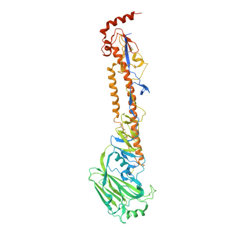 | |
UniProt | |||||
Entity Groups | |||||
| Sequence Clusters | 30% Identity50% Identity70% Identity90% Identity95% Identity100% Identity | ||||
| UniProt Group | A0A059VBQ9 | ||||
Sequence AnnotationsExpand | |||||
Reference Sequence | |||||
| Ligands 1 Unique | |||||
|---|---|---|---|---|---|
| ID | Chains | Name / Formula / InChI Key | 2D Diagram | 3D Interactions | |
| SIA Download:Ideal Coordinates CCD File | B [auth A] | N-acetyl-alpha-neuraminic acid C11 H19 N O9 SQVRNKJHWKZAKO-YRMXFSIDSA-N |  | ||
| Length ( Å ) | Angle ( ˚ ) |
|---|---|
| a = 110.306 | α = 90 |
| b = 110.306 | β = 90 |
| c = 314.111 | γ = 120 |
| Software Name | Purpose |
|---|---|
| PHENIX | refinement |
| HKL-2000 | data reduction |
| HKL-2000 | data scaling |
| Coot | model building |
| PHASER | phasing |


