6II1 | pdb_00006ii1

Crystal Structure Analysis of CO form hemoglobin from Bos taurus


Experimental Data Snapshot

  • Method: X-RAY DIFFRACTION
  • Resolution: 1.34 Å
  • R-Value Free: 
    0.175 (Depositor), 0.184 (DCC) 
  • R-Value Work: 
    0.137 (Depositor), 0.147 (DCC) 
  • R-Value Observed: 
    0.138 (Depositor) 

Starting Model: experimental
View more details

wwPDB Validation 3D Report Full Report

Validation slider image for 6II1

Ligand Structure Quality Assessment 


This is version 1.2 of the entry. See complete history

Literature

Quaternary Structure Analysis of a Hemoglobin Core in Hemoglobin-Albumin Cluster.

Morita, Y.Yamada, T.Kureishi, M.Kihira, K.Komatsu, T.

(2018) J Phys Chem B 122: 12031-12039

  • DOI: https://doi.org/10.1021/acs.jpcb.8b10077
  • Primary Citation Related Structures: 
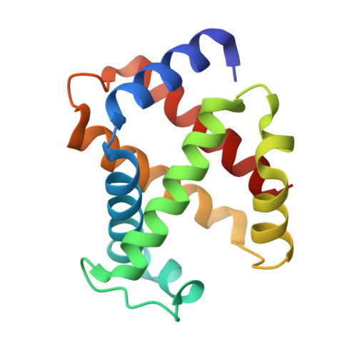
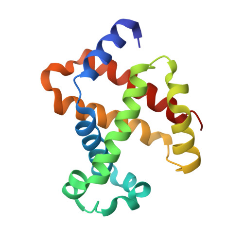
    6IHX, 6II1

  • PubMed Abstract: 

    A core-shell ensemble of bovine hemoglobin (Hb) and human serum albumin (HSA) is an artificial O 2 carrier as a red blood cell substitute. This protein particle is created by covalent wrapping of a carbonyl Hb with HSAs: Hb R -HSA 3 cluster, where Hb R signifies the use of carbonyl Hb (relaxed (R) state conformation) as a starting material. The Hb R -HSA 3 cluster exhibits high O 2 affinity and low cooperativity. Analysis of the quaternary structure of the central Hb R in the cluster revealed that its high O 2 affinity is attributed to the physically immobile Hb R nucleus. Circular dichroism and UV-vis absorption spectroscopy showed that the structure of deoxy Hb R core closely resembles the R-state. The crystal structure of Lys-modified carbonyl Hb R was superimposed on that of carbonyl Hb. These results imply that chemical modifications of the surface Lys groups and Cys-93(β) of the carbonyl Hb with cross-linking agent interfered in the quaternary structure movement from the R-state to tense (T) state. As expected, coupling of deoxy Hb (T-state) with HSAs yielded Hb T -HSA 3 cluster having low O 2 affinity. The mixing of Hb R -HSA 3 and Hb T -HSA 3 clusters conferred a tailor-made formulation of artificial O 2 carrier with a desired O 2 affinity ( P 50 ).


  • Organizational Affiliation
    • Department of Applied Chemistry, Faculty of Science and Engineering , Chuo University , 1-13-27 Kasuga , Bunkyo-ku , Tokyo 112-8551 , Japan.

Macromolecule Content 

  • Total Structure Weight: 63.79 kDa 
  • Atom Count: 5,230 
  • Modeled Residue Count: 563 
  • Deposited Residue Count: 566 
  • Unique protein chains: 2

Macromolecules

Find similar proteins by:|  3D Structure
Entity ID: 1
MoleculeChains  Sequence LengthOrganismDetailsImage
Hemoglobin subunit alpha
A, C
138Bos taurusMutation(s): 0 
UniProt
Find proteins for P01966 (Bos taurus)
Explore P01966 
Go to UniProtKB:  P01966
Entity Groups
Sequence Clusters30% Identity50% Identity70% Identity90% Identity95% Identity100% Identity
UniProt GroupP01966
Sequence Annotations
Expand
Reference Sequence
Find similar proteins by:|  3D Structure
Entity ID: 2
MoleculeChains  Sequence LengthOrganismDetailsImage
Hemoglobin subunit beta
B, D
145Bos taurusMutation(s): 0 
UniProt
Find proteins for P02070 (Bos taurus)
Explore P02070 
Go to UniProtKB:  P02070
Entity Groups
Sequence Clusters30% Identity50% Identity70% Identity90% Identity95% Identity100% Identity
UniProt GroupP02070
Sequence Annotations
Expand
Reference Sequence

Small Molecules

Ligands 2 Unique
IDChains Name / Formula / InChI Key2D Diagram3D Interactions
HEM

Query on HEM


Download:Ideal Coordinates CCD File
E [auth A],
G [auth B],
I [auth C],
K [auth D]
PROTOPORPHYRIN IX CONTAINING FE
C34 H32 Fe N4 O4
KABFMIBPWCXCRK-RGGAHWMASA-L
CMO

Query on CMO


Download:Ideal Coordinates CCD File
F [auth A],
H [auth B],
J [auth C],
L [auth D]
CARBON MONOXIDE
C O
UGFAIRIUMAVXCW-UHFFFAOYSA-N

Experimental Data & Validation

Experimental Data

  • Method: X-RAY DIFFRACTION
  • Resolution: 1.34 Å
  • R-Value Free:  0.175 (Depositor), 0.184 (DCC) 
  • R-Value Work:  0.137 (Depositor), 0.147 (DCC) 
  • R-Value Observed: 0.138 (Depositor) 
Space Group: P 21 21 21
Unit Cell:
Length ( Å )Angle ( ˚ )
a = 63.56α = 90
b = 76.283β = 90
c = 109.789γ = 90
Software Package:
Software NamePurpose
XDSdata reduction
Aimlessdata scaling
PHASERphasing
REFMACrefinement
PDB_EXTRACTdata extraction

Structure Validation

View Full Validation Report


Ligand Structure Quality Assessment 


Entry History 

Deposition Data

Revision History  (Full details and data files)

  • Version 1.0: 2018-12-12
    Type: Initial release
  • Version 1.1: 2019-04-24
    Changes: Data collection, Database references
  • Version 1.2: 2023-11-22
    Changes: Data collection, Database references, Refinement description