6IEM | pdb_00006iem

Argininosuccinate lyase from Mycobacterium tuberculosis


Experimental Data Snapshot

  • Method: X-RAY DIFFRACTION
  • Resolution: 2.20 Å
  • R-Value Free: 
    0.244 (Depositor), 0.242 (DCC) 
  • R-Value Work: 
    0.203 (Depositor), 0.203 (DCC) 
  • R-Value Observed: 
    0.205 (Depositor) 

Starting Model: experimental
View more details

wwPDB Validation 3D Report Full Report

Validation slider image for 6IEM

Ligand Structure Quality Assessment 


This is version 1.2 of the entry. See complete history

Literature

Structural studies on M. tuberculosis argininosuccinate lyase and its liganded complex: Insights into catalytic mechanism.

Paul, A.Mishra, A.Surolia, A.Vijayan, M.

(2019) IUBMB Life 71: 643-652

  • DOI: https://doi.org/10.1002/iub.2000
  • Primary Citation Related Structures: 
    6IEM, 6IEN

  • PubMed Abstract: 

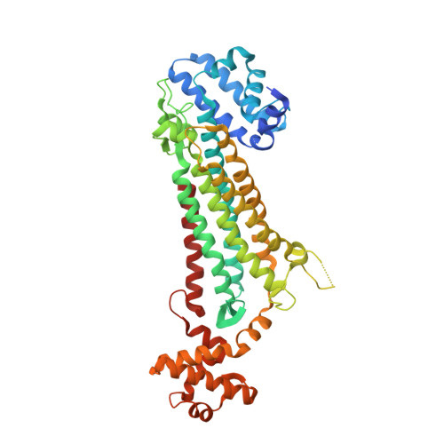
    Argininosuccinate lyase catalyses the reversible breakdown of argininosuccinate into arginine and fumarate and is known to form tetramers in its quaternary association. The absence of structures involving competent enzymes bound to substrate/products came in the way of the precise elucidation of the catalytic mechanism of this family of proteins. Crystal structures of the enzyme from Mycobacterium tuberculosis in an unliganded form and its complex with the substrate/products have now been determined at 2.2 and 2.7 Å, respectively. The refinement of the structure of the complex was bedevilled by the presence of a lattice translocation defect. The two tetramers in the apo-crystals and the one in the crystals of the liganded protein, have the same structure except for the movements associated with enzyme action. Each molecule consists of an N-domain, an M-domain, and a C-domain. The molecule consists of four binding sites, each made up of peptide stretches from three subunits. Three binding sites appear to be occupied by the ligand in the transition state, while the products occupy the fourth site. The structure exhibits the movement of a loop in the M-domain and parts of the C-domain. This is the first instance when the appropriate movements are observed in a complex with bound substrate/product. The detailed picture of the binding site, active site residues and the movements associated with catalysis thus obtained, enabled a revisit of the mechanism of action of the enzyme. © 2019 IUBMB Life, 71(5):643-652, 2019.


  • Organizational Affiliation
    • Molecular Biophysics Unit, Indian Institute of Science, Bangalore, India.

Macromolecule Content 

  • Total Structure Weight: 400.45 kDa 
  • Atom Count: 27,041 
  • Modeled Residue Count: 3,617 
  • Deposited Residue Count: 3,760 
  • Unique protein chains: 1

Macromolecules

Find similar proteins by:|  3D Structure
Entity ID: 1
MoleculeChains  Sequence LengthOrganismDetailsImage
Argininosuccinate lyase
A, B, C, D, E
A, B, C, D, E, F, G, H
470Mycobacterium tuberculosis H37RvMutation(s): 0 
Gene Names: argHRv1659MTCY06H11.24
EC: 4.3.2.1
UniProt
Find proteins for P9WPY7 (Mycobacterium tuberculosis (strain ATCC 25618 / H37Rv))
Explore P9WPY7 
Go to UniProtKB:  P9WPY7
Entity Groups
Sequence Clusters30% Identity50% Identity70% Identity90% Identity95% Identity100% Identity
UniProt GroupP9WPY7
Sequence Annotations
Expand
Reference Sequence

Small Molecules

Ligands 6 Unique
IDChains Name / Formula / InChI Key2D Diagram3D Interactions
2PE

Query on 2PE


Download:Ideal Coordinates CCD File
S [auth E]NONAETHYLENE GLYCOL
C18 H38 O10
YZUUTMGDONTGTN-UHFFFAOYSA-N
P3G

Query on P3G


Download:Ideal Coordinates CCD File
R [auth D]3,6,9,12,15-PENTAOXAHEPTADECANE
C12 H26 O5
HYDWALOBQJFOMS-UHFFFAOYSA-N
PG4

Query on PG4


Download:Ideal Coordinates CCD File
K [auth B],
N [auth C]
TETRAETHYLENE GLYCOL
C8 H18 O5
UWHCKJMYHZGTIT-UHFFFAOYSA-N
PGE

Query on PGE


Download:Ideal Coordinates CCD File
U [auth F]TRIETHYLENE GLYCOL
C6 H14 O4
ZIBGPFATKBEMQZ-UHFFFAOYSA-N
GOL

Query on GOL


Download:Ideal Coordinates CCD File
I [auth A],
L [auth B]
GLYCEROL
C3 H8 O3
PEDCQBHIVMGVHV-UHFFFAOYSA-N
EDO

Query on EDO


Download:Ideal Coordinates CCD File
J [auth A]
M [auth B]
O [auth C]
P [auth D]
Q [auth D]
J [auth A],
M [auth B],
O [auth C],
P [auth D],
Q [auth D],
T [auth E],
V [auth F]
1,2-ETHANEDIOL
C2 H6 O2
LYCAIKOWRPUZTN-UHFFFAOYSA-N

Experimental Data & Validation

Experimental Data

  • Method: X-RAY DIFFRACTION
  • Resolution: 2.20 Å
  • R-Value Free:  0.244 (Depositor), 0.242 (DCC) 
  • R-Value Work:  0.203 (Depositor), 0.203 (DCC) 
  • R-Value Observed: 0.205 (Depositor) 
Space Group: P 21 21 21
Unit Cell:
Length ( Å )Angle ( ˚ )
a = 127.55α = 90
b = 183.82β = 90
c = 190.6γ = 90
Software Package:
Software NamePurpose
REFMACrefinement
MOSFLMdata reduction
SCALAdata scaling
PHASERphasing

Structure Validation

View Full Validation Report


Ligand Structure Quality Assessment 


Entry History 

Deposition Data

Revision History  (Full details and data files)

  • Version 1.0: 2019-02-20
    Type: Initial release
  • Version 1.1: 2019-04-17
    Changes: Data collection, Database references
  • Version 1.2: 2023-11-22
    Changes: Data collection, Database references, Refinement description