6ICP | pdb_00006icp

Pseudomonas putida CBB5 NdmA QL mutant with caffeine


Experimental Data Snapshot

  • Method: X-RAY DIFFRACTION
  • Resolution: 2.20 Å
  • R-Value Free: 
    0.221 (Depositor), 0.221 (DCC) 
  • R-Value Work: 
    0.179 (Depositor), 0.179 (DCC) 
  • R-Value Observed: 
    0.180 (Depositor) 

wwPDB Validation 3D Report Full Report

Validation slider image for 6ICP

Ligand Structure Quality Assessment 


This is version 1.2 of the entry. See complete history

Literature

Structural and Mechanistic Insights into Caffeine Degradation by the Bacterial N-Demethylase Complex.

Kim, J.H.Kim, B.H.Brooks, S.Kang, S.Y.Summers, R.M.Song, H.K.

(2019) J Mol Biology 431: 3647-3661

  • DOI: https://doi.org/10.1016/j.jmb.2019.08.004
  • Primary Citation Related Structures: 
    6ICK, 6ICL, 6ICM, 6ICN, 6ICO, 6ICP, 6ICQ

  • PubMed Abstract: 

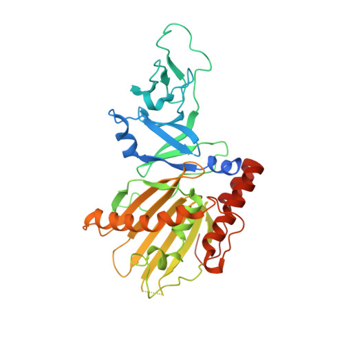
    Caffeine, found in many foods, beverages, and pharmaceuticals, is the most used chemical compound for mental alertness. It is originally a natural product of plants and exists widely in environmental soil. Some bacteria, such as Pseudomonas putida CBB5, utilize caffeine as a sole carbon and nitrogen source by degrading it through sequential N-demethylation catalyzed by five enzymes (NdmA, NdmB, NdmC, NdmD, and NdmE). The environmentally friendly enzymatic reaction products, methylxanthines, are high-value biochemicals that are used in the pharmaceutical and cosmetic industries. However, the structures and biochemical properties of bacterial N-demethylases remain largely unknown. Here, we report the structures of NdmA and NdmB, the initial N 1 - and N 3 -specific demethylases, respectively. Reverse-oriented substrate bindings were observed in the substrate-complexed structures, offering methyl position specificity for proper N-demethylation. For efficient sequential degradation of caffeine, these enzymes form a unique heterocomplex with 3:3 stoichiometry, which was confirmed by enzymatic assays, fluorescent labeling, and small-angle x-ray scattering. The binary structure of NdmA with the ferredoxin domain of NdmD, which is the first structural information for the plant-type ferredoxin domain in a complex state, was also determined to better understand electron transport during N-demethylation. These findings broaden our understanding of the caffeine degradation mechanism by bacterial enzymes and will enable their use for industrial applications.


  • Organizational Affiliation
    • Department of Life Sciences, Korea University, 145 Anam-ro, Seongbuk-gu, Seoul 02841, Republic of Korea.

Macromolecule Content 

  • Total Structure Weight: 128.36 kDa 
  • Atom Count: 8,804 
  • Modeled Residue Count: 1,003 
  • Deposited Residue Count: 1,107 
  • Unique protein chains: 1

Macromolecules

Find similar proteins by:|  3D Structure
Entity ID: 1
MoleculeChains  Sequence LengthOrganismDetailsImage
Methylxanthine N1-demethylase NdmA
A, B, C
369Pseudomonas putidaMutation(s): 2 
Gene Names: ndmA
EC: 1.14.13.178
UniProt
Find proteins for H9N289 (Pseudomonas putida)
Explore H9N289 
Go to UniProtKB:  H9N289
Entity Groups
Sequence Clusters30% Identity50% Identity70% Identity90% Identity95% Identity100% Identity
UniProt GroupH9N289
Sequence Annotations
Expand
Reference Sequence

Small Molecules

Ligands 3 Unique
IDChains Name / Formula / InChI Key2D Diagram3D Interactions
CFF

Query on CFF


Download:Ideal Coordinates CCD File
F [auth A],
I [auth B],
L [auth C]
CAFFEINE
C8 H10 N4 O2
RYYVLZVUVIJVGH-UHFFFAOYSA-N
FES

Query on FES


Download:Ideal Coordinates CCD File
D [auth A],
G [auth B],
J [auth C]
FE2/S2 (INORGANIC) CLUSTER
Fe2 S2
NIXDOXVAJZFRNF-UHFFFAOYSA-N
FE

Query on FE


Download:Ideal Coordinates CCD File
E [auth A],
H [auth B],
K [auth C]
FE (III) ION
Fe
VTLYFUHAOXGGBS-UHFFFAOYSA-N

Experimental Data & Validation

Experimental Data

  • Method: X-RAY DIFFRACTION
  • Resolution: 2.20 Å
  • R-Value Free:  0.221 (Depositor), 0.221 (DCC) 
  • R-Value Work:  0.179 (Depositor), 0.179 (DCC) 
  • R-Value Observed: 0.180 (Depositor) 
Space Group: P 32 2 1
Unit Cell:
Length ( Å )Angle ( ˚ )
a = 136.243α = 90
b = 136.243β = 90
c = 154.087γ = 120
Software Package:
Software NamePurpose
PHENIXrefinement
HKL-2000data reduction
HKL-2000data scaling
PHENIXphasing

Structure Validation

View Full Validation Report


Ligand Structure Quality Assessment 


Entry History 

& Funding Information

Deposition Data


Funding OrganizationLocationGrant Number
National Research Foundation (Korea)Korea, Republic Of2016R1E1A1A01942623

Revision History  (Full details and data files)

  • Version 1.0: 2019-09-04
    Type: Initial release
  • Version 1.1: 2019-09-25
    Changes: Data collection, Database references
  • Version 1.2: 2024-03-27
    Changes: Data collection, Database references, Derived calculations