6IBL

ACTIVATED TURKEY BETA1 ADRENOCEPTOR WITH BOUND AGONIST FORMOTEROL AND NANOBODY Nb80


Experimental Data Snapshot

  • Method: X-RAY DIFFRACTION
  • Resolution: 2.70 Å
  • R-Value Free: 0.277 
  • R-Value Work: 0.240 
  • R-Value Observed: 0.241 

Starting Models: experimental
View more details

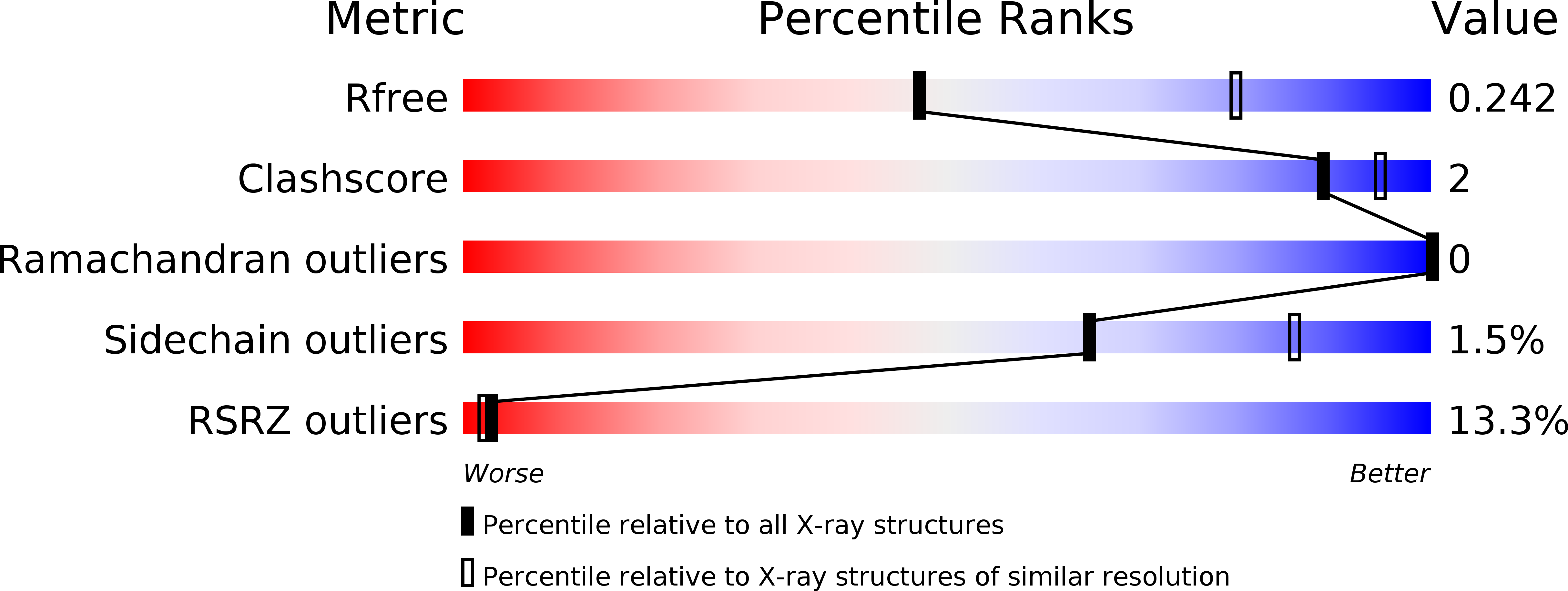
wwPDB Validation   3D Report Full Report


Ligand Structure Quality Assessment 


This is version 1.5 of the entry. See complete history


Literature

Molecular basis of beta-arrestin coupling to formoterol-bound beta1-adrenoceptor.

Lee, Y.Warne, T.Nehme, R.Pandey, S.Dwivedi-Agnihotri, H.Chaturvedi, M.Edwards, P.C.Garcia-Nafria, J.Leslie, A.G.W.Shukla, A.K.Tate, C.G.

(2020) Nature 583: 862-866

  • DOI: https://doi.org/10.1038/s41586-020-2419-1
  • Primary Citation of Related Structures:  
    6IBL, 6TKO

  • PubMed Abstract: 

    The β 1 -adrenoceptor (β 1 AR) is a G-protein-coupled receptor (GPCR) that couples 1 to the heterotrimeric G protein G s . G-protein-mediated signalling is terminated by phosphorylation of the C terminus of the receptor by GPCR kinases (GRKs) and by coupling of β-arrestin 1 (βarr1, also known as arrestin 2), which displaces G s and induces signalling through the MAP kinase pathway 2 . The ability of synthetic agonists to induce signalling preferentially through either G proteins or arrestins-known as biased agonism 3 -is important in drug development, because the therapeutic effect may arise from only one signalling cascade, whereas the other pathway may mediate undesirable side effects 4 . To understand the molecular basis for arrestin coupling, here we determined the cryo-electron microscopy structure of the β 1 AR-βarr1 complex in lipid nanodiscs bound to the biased agonist formoterol 5 , and the crystal structure of formoterol-bound β 1 AR coupled to the G-protein-mimetic nanobody 6 Nb80. βarr1 couples to β 1 AR in a manner distinct to that 7 of G s coupling to β 2 AR-the finger loop of βarr1 occupies a narrower cleft on the intracellular surface, and is closer to transmembrane helix H7 of the receptor when compared with the C-terminal α5 helix of G s . The conformation of the finger loop in βarr1 is different from that adopted by the finger loop of visual arrestin when it couples to rhodopsin 8 . β 1 AR coupled to βarr1 shows considerable differences in structure compared with β 1 AR coupled to Nb80, including an inward movement of extracellular loop 3 and the cytoplasmic ends of H5 and H6. We observe weakened interactions between formoterol and two serine residues in H5 at the orthosteric binding site of β 1 AR, and find that formoterol has a lower affinity for the β 1 AR-βarr1 complex than for the β 1 AR-G s complex. The structural differences between these complexes of β 1 AR provide a foundation for the design of small molecules that could bias signalling in the β-adrenoceptors.


  • Organizational Affiliation

    MRC Laboratory of Molecular Biology, Cambridge, UK.


Macromolecules
Find similar proteins by:  (by identity cutoff)  |  3D Structure
Entity ID: 1
MoleculeChains Sequence LengthOrganismDetailsImage
Thioredoxin 1,Beta-1 adrenergic receptorA,
C [auth B]
416Escherichia coli K-12Meleagris gallopavo
This entity is chimeric
Mutation(s): 6 
Gene Names: trxAfipAtsnCb3781JW5856ADRB1
Membrane Entity: Yes 
UniProt
Find proteins for P07700 (Meleagris gallopavo)
Explore P07700 
Go to UniProtKB:  P07700
Find proteins for P0AA25 (Escherichia coli (strain K12))
Explore P0AA25 
Go to UniProtKB:  P0AA25
Entity Groups  
Sequence Clusters30% Identity50% Identity70% Identity90% Identity95% Identity100% Identity
UniProt GroupsP07700P0AA25
Sequence Annotations
Expand
  • Reference Sequence
Find similar proteins by:  (by identity cutoff)  |  3D Structure
Entity ID: 2
MoleculeChains Sequence LengthOrganismDetailsImage
Camelid antibody fragment Nb80B [auth C],
D
121Lama glamaMutation(s): 0 
Entity Groups  
Sequence Clusters30% Identity50% Identity70% Identity90% Identity95% Identity100% Identity
Sequence Annotations
Expand
  • Reference Sequence
Small Molecules
Ligands 3 Unique
IDChains Name / Formula / InChI Key2D Diagram3D Interactions
2CV
Query on 2CV

Download Ideal Coordinates CCD File 
G [auth A]
H [auth A]
I [auth A]
J [auth A]
K [auth A]
G [auth A],
H [auth A],
I [auth A],
J [auth A],
K [auth A],
N [auth B],
O [auth B],
P [auth B]
HEGA-10
C18 H37 N O7
ITEIKACYSCODFV-ATLSCFEFSA-N
H98
Query on H98

Download Ideal Coordinates CCD File 
E [auth A],
L [auth B]
~{N}-[5-[(1~{R})-2-[[(2~{R})-1-(4-methoxyphenyl)propan-2-yl]amino]-1-oxidanyl-ethyl]-2-oxidanyl-phenyl]methanamide
C19 H24 N2 O4
BPZSYCZIITTYBL-YJYMSZOUSA-N
NA
Query on NA

Download Ideal Coordinates CCD File 
F [auth A],
M [auth B]
SODIUM ION
Na
FKNQFGJONOIPTF-UHFFFAOYSA-N
Experimental Data & Validation

Experimental Data

  • Method: X-RAY DIFFRACTION
  • Resolution: 2.70 Å
  • R-Value Free: 0.277 
  • R-Value Work: 0.240 
  • R-Value Observed: 0.241 
  • Space Group: P 21 21 21
Unit Cell:
Length ( Å )Angle ( ˚ )
a = 116.65α = 90
b = 121.12β = 90
c = 129.84γ = 90
Software Package:
Software NamePurpose
REFMACrefinement
MOSFLMdata reduction
Aimlessdata scaling
REFMACphasing

Structure Validation

View Full Validation Report


Ligand Structure Quality Assessment 


Entry History 

Deposition Data

Revision History  (Full details and data files)

  • Version 1.0: 2019-01-09
    Type: Initial release
  • Version 1.1: 2019-05-22
    Changes: Data collection, Database references
  • Version 1.2: 2020-07-08
    Changes: Database references
  • Version 1.3: 2020-08-12
    Changes: Database references, Derived calculations
  • Version 1.4: 2024-01-24
    Changes: Data collection, Database references, Refinement description
  • Version 1.5: 2024-11-20
    Changes: Structure summary