6IB0 | pdb_00006ib0

The structure of MKK7 in complex with the covalent 4-amino-pyrazolopyrimidine 3a


Experimental Data Snapshot

  • Method: X-RAY DIFFRACTION
  • Resolution: 2.60 Å
  • R-Value Free: 
    0.271 (Depositor), 0.277 (DCC) 
  • R-Value Work: 
    0.227 (Depositor), 0.231 (DCC) 
  • R-Value Observed: 
    0.230 (Depositor) 

Starting Model: experimental
View more details

wwPDB Validation 3D Report Full Report

Validation slider image for 6IB0

Ligand Structure Quality Assessment 


This is version 1.3 of the entry. See complete history

Literature

Targeting the MKK7-JNK (Mitogen-Activated Protein Kinase Kinase 7-c-Jun N-Terminal Kinase) Pathway with Covalent Inhibitors.

Wolle, P.Hardick, J.Cronin, S.J.F.Engel, J.Baumann, M.Lategahn, J.Penninger, J.M.Rauh, D.

(2019) J Med Chem 62: 2843-2848

  • DOI: https://doi.org/10.1021/acs.jmedchem.9b00102
  • Primary Citation Related Structures: 
    6IB0, 6IB2

  • PubMed Abstract: 

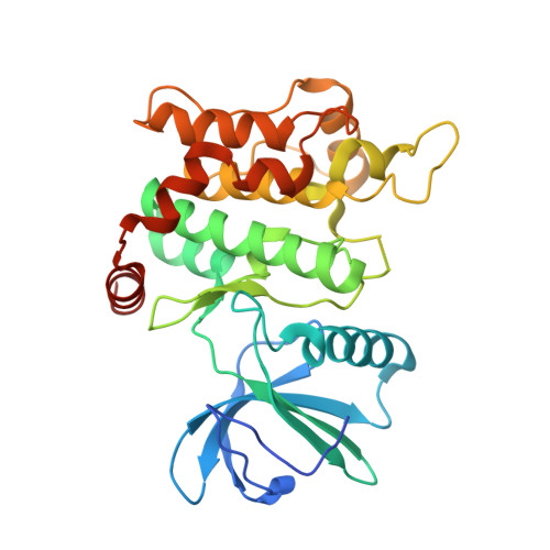
    The protein kinase MKK7 is linked to neuronal development and the onset of cancer. The field, however, lacks high-quality functional probes that would allow for the dissection of its detailed functions. Against this background, we describe an effective covalent inhibitor of MKK7 based on the pyrazolopyrimidine scaffold.


  • Organizational Affiliation
    • Faculty of Chemistry and Chemical Biology , TU Dortmund University , Otto-Hahn-Strasse 4a , 44227 Dortmund , Germany.

Macromolecule Content 

  • Total Structure Weight: 36.64 kDa 
  • Atom Count: 2,323 
  • Modeled Residue Count: 290 
  • Deposited Residue Count: 318 
  • Unique protein chains: 1

Macromolecules

Find similar proteins by:|  3D Structure
Entity ID: 1
MoleculeChains  Sequence LengthOrganismDetailsImage
Dual specificity mitogen-activated protein kinase kinase 7318Homo sapiensMutation(s): 0 
Gene Names: MAP2K7JNKK2MEK7MKK7PRKMK7SKK4
EC: 2.7.12.2
UniProt & NIH Common Fund Data Resources
Find proteins for O14733 (Homo sapiens)
Explore O14733 
Go to UniProtKB:  O14733
PHAROS:  O14733
GTEx:  ENSG00000076984 
Entity Groups
Sequence Clusters30% Identity50% Identity70% Identity90% Identity95% Identity100% Identity
UniProt GroupO14733
Sequence Annotations
Expand
Reference Sequence

Small Molecules

Ligands 2 Unique
IDChains Name / Formula / InChI Key2D Diagram3D Interactions
H8Z

Query on H8Z


Download:Ideal Coordinates CCD File
B [auth A]1-[(3~{R})-3-(4-azanyl-3-ethynyl-pyrazolo[3,4-d]pyrimidin-1-yl)piperidin-1-yl]prop-2-en-1-one
C15 H16 N6 O
XXFDMGXYEIKFNZ-SNVBAGLBSA-N
PG4

Query on PG4


Download:Ideal Coordinates CCD File
C [auth A]TETRAETHYLENE GLYCOL
C8 H18 O5
UWHCKJMYHZGTIT-UHFFFAOYSA-N
Binding Affinity Annotations 
IDSourceBinding Affinity
H8Z BindingDB:  6IB0 IC50: 30 (nM) from 1 assay(s)
EC50: 1.00e+4 (nM) from 1 assay(s)

Experimental Data & Validation

Experimental Data

  • Method: X-RAY DIFFRACTION
  • Resolution: 2.60 Å
  • R-Value Free:  0.271 (Depositor), 0.277 (DCC) 
  • R-Value Work:  0.227 (Depositor), 0.231 (DCC) 
  • R-Value Observed: 0.230 (Depositor) 
Space Group: P 21 21 21
Unit Cell:
Length ( Å )Angle ( ˚ )
a = 59.4α = 90
b = 66β = 90
c = 86.4γ = 90
Software Package:
Software NamePurpose
PHENIXrefinement
XDSdata reduction
XSCALEdata scaling
PHENIXphasing

Structure Validation

View Full Validation Report


Ligand Structure Quality Assessment 


Entry History 

Deposition Data

Revision History  (Full details and data files)

  • Version 1.0: 2019-02-27
    Type: Initial release
  • Version 1.1: 2019-03-27
    Changes: Data collection, Database references
  • Version 1.2: 2024-01-24
    Changes: Data collection, Database references, Refinement description
  • Version 1.3: 2024-10-23
    Changes: Structure summary