6IAI | pdb_00006iai

StoD is a novel Salmonella Typhi type III secretion system E3 ubiquitin ligase effector


Experimental Data Snapshot

  • Method: X-RAY DIFFRACTION
  • Resolution: 2.54 Å
  • R-Value Free: 
    0.244 (Depositor), 0.249 (DCC) 
  • R-Value Work: 
    0.216 (Depositor), 0.212 (DCC) 
  • R-Value Observed: 
    0.217 (Depositor) 

wwPDB Validation 3D Report Full Report

Validation slider image for 6IAI

This is version 1.2 of the entry. See complete history

Literature

The S . Typhi effector StoD is an E3/E4 ubiquitin ligase which binds K48- and K63-linked diubiquitin.

McDowell, M.A.Byrne, A.M.Mylona, E.Johnson, R.Sagfors, A.Crepin, V.F.Lea, S.Frankel, G.

(2019) Life Sci Alliance 2

  • DOI: https://doi.org/10.26508/lsa.201800272
  • Primary Citation Related Structures: 
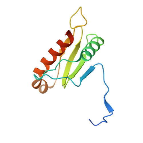
    6IAI

  • PubMed Abstract: 

    Salmonella enterica (e.g., serovars Typhi and Typhimurium) relies on translocation of effectors via type III secretion systems (T3SS). Specialization of typhoidal serovars is thought to be mediated via pseudogenesis. Here, we show that the Salmonella Typhi STY1076/t1865 protein, named StoD, a homologue of the enteropathogenic Escherichia coli /enterohemorrhagic E. coli/Citrobacter rodentium NleG, is a T3SS effector. The StoD C terminus (StoD-C) is a U-box E3 ubiquitin ligase, capable of autoubiquitination in the presence of multiple E2s. The crystal structure of the StoD N terminus (StoD-N) at 2.5 Å resolution revealed a ubiquitin-like fold. In HeLa cells expressing StoD, ubiquitin is redistributed into puncta that colocalize with StoD. Binding assays showed that StoD-N and StoD-C bind the same exposed surface of the β-sheet of ubiquitin, suggesting that StoD could simultaneously interact with two ubiquitin molecules. Consistently, StoD interacted with both K63- (K D = 5.6 ± 1 μM) and K48-linked diubiquitin (K D = 15 ± 4 μM). Accordingly, we report the first S. Typhi-specific T3SS effector. We suggest that StoD recognizes and ubiquitinates pre-ubiquitinated targets, thus subverting intracellular signaling by functioning as an E4 enzyme.


  • Organizational Affiliation
    • Sir William Dunn School of Pathology, University of Oxford, Oxford, UK.

Macromolecule Content 

  • Total Structure Weight: 55.59 kDa 
  • Atom Count: 3,530 
  • Modeled Residue Count: 429 
  • Deposited Residue Count: 484 
  • Unique protein chains: 1

Macromolecules

Find similar proteins by:|  3D Structure
Entity ID: 1
MoleculeChains  Sequence LengthOrganismDetailsImage
Protein of uncharacterized function (DUF1076)
A, B, C, D
121Salmonella enterica subsp. enterica serovar TyphiMutation(s): 0 
Gene Names: STY1076t1865NCTC8385_01041
UniProt
Find proteins for Q8Z7T2 (Salmonella typhi)
Explore Q8Z7T2 
Go to UniProtKB:  Q8Z7T2
Entity Groups
Sequence Clusters30% Identity50% Identity70% Identity90% Identity95% Identity100% Identity
UniProt GroupQ8Z7T2
Sequence Annotations
Expand
Reference Sequence

Experimental Data & Validation

Experimental Data

  • Method: X-RAY DIFFRACTION
  • Resolution: 2.54 Å
  • R-Value Free:  0.244 (Depositor), 0.249 (DCC) 
  • R-Value Work:  0.216 (Depositor), 0.212 (DCC) 
  • R-Value Observed: 0.217 (Depositor) 
Space Group: P 43 2 2
Unit Cell:
Length ( Å )Angle ( ˚ )
a = 92.96α = 90
b = 92.96β = 90
c = 156γ = 90
Software Package:
Software NamePurpose
BUSTERrefinement
xia2data reduction
XSCALEdata scaling
SHARPphasing

Structure Validation

View Full Validation Report


Entry History 

& Funding Information

Deposition Data


Funding OrganizationLocationGrant Number
Wellcome TrustUnited Kingdom100298/Z/12/Z
Wellcome TrustUnited Kingdom107057/Z/15/Z
Biotechnology and Biological Sciences Research CouncilUnited KingdomBB/K001515/1

Revision History  (Full details and data files)

  • Version 1.0: 2019-05-29
    Type: Initial release
  • Version 1.1: 2020-12-09
    Changes: Database references
  • Version 1.2: 2024-05-15
    Changes: Data collection, Database references