6I8C | pdb_00006i8c

Crystal structure of the murine beta-2-microglobulin.


Experimental Data Snapshot

  • Method: X-RAY DIFFRACTION
  • Resolution: 1.92 Å
  • R-Value Free: 
    0.222 (Depositor), 0.217 (DCC) 
  • R-Value Work: 
    0.183 (Depositor), 0.195 (DCC) 
  • R-Value Observed: 
    0.185 (Depositor) 

Starting Model: experimental
View more details

wwPDB Validation 3D Report Full Report

Validation slider image for 6I8C

This is version 2.3 of the entry. See complete history

Literature

Biochemical and biophysical comparison of human and mouse beta-2 microglobulin reveals the molecular determinants of low amyloid propensity.

Achour, A.Broggini, L.Han, X.Sun, R.Santambrogio, C.Buratto, J.Visentin, C.Barbiroli, A.De Luca, C.M.G.Sormanni, P.Moda, F.De Simone, A.Sandalova, T.Grandori, R.Camilloni, C.Ricagno, S.

(2020) FEBS J 287: 546-560

  • DOI: https://doi.org/10.1111/febs.15046
  • Primary Citation Related Structures: 
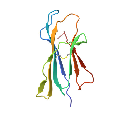
    6I8C

  • PubMed Abstract: 

    The molecular bases of amyloid aggregation propensity are still poorly understood, especially for proteins that display a stable folded native structure. A prototypic example is human beta-2 microglobulin (β2m), which, when accumulated in patients, gives rise to dialysis-related amyloidosis. Interestingly, although the physiologic concentration of β2m in mice is five times higher than that found in human patients, no amyloid deposits are observed in mice. Moreover, murine β2m (mβ2m) not only displays a lower amyloid propensity both in vivo and in vitro but also inhibits the aggregation of human β2m in vitro. Here, we compared human and mβ2m for their aggregation propensity, ability to form soluble oligomers, stability, three-dimensional structure and dynamics. Our results indicate that mβ2m low-aggregation propensity is due to two concomitant aspects: the low-aggregation propensity of its primary sequence combined with the absence of high-energy amyloid-competent conformations under native conditions. The identification of the specific properties determining the low-aggregation propensity of mouse β2m will help delineate the molecular risk factors which cause a folded protein to aggregate.


  • Organizational Affiliation
    • Science for Life Laboratory, Department of Medicine Solna, Karolinska Institute, Solna, Sweden.

Macromolecule Content 

  • Total Structure Weight: 11.85 kDa 
  • Atom Count: 925 
  • Modeled Residue Count: 99 
  • Deposited Residue Count: 99 
  • Unique protein chains: 1

Macromolecules

Find similar proteins by:|  3D Structure
Entity ID: 1
MoleculeChains  Sequence LengthOrganismDetailsImage
Beta-2-microglobulin99Mus musculusMutation(s): 1 
Gene Names: B2m
UniProt & NIH Common Fund Data Resources
Find proteins for P01887 (Mus musculus)
Explore P01887 
Go to UniProtKB:  P01887
IMPC:  MGI:88127
Entity Groups
Sequence Clusters30% Identity50% Identity70% Identity90% Identity95% Identity100% Identity
UniProt GroupP01887
Sequence Annotations
Expand
Reference Sequence

Small Molecules

Ligands 1 Unique
IDChains Name / Formula / InChI Key2D Diagram3D Interactions
PGE

Query on PGE


Download:Ideal Coordinates CCD File
B [auth A]TRIETHYLENE GLYCOL
C6 H14 O4
ZIBGPFATKBEMQZ-UHFFFAOYSA-N

Experimental Data & Validation

Experimental Data

  • Method: X-RAY DIFFRACTION
  • Resolution: 1.92 Å
  • R-Value Free:  0.222 (Depositor), 0.217 (DCC) 
  • R-Value Work:  0.183 (Depositor), 0.195 (DCC) 
  • R-Value Observed: 0.185 (Depositor) 
Space Group: P 21 21 21
Unit Cell:
Length ( Å )Angle ( ˚ )
a = 36.242α = 90
b = 47.603β = 90
c = 64.937γ = 90
Software Package:
Software NamePurpose
REFMACrefinement
DENZOdata reduction
SCALEPACKdata scaling
AMoREphasing

Structure Validation

View Full Validation Report


Entry History 

Deposition Data

Revision History  (Full details and data files)

  • Version 1.0: 2019-10-02
    Type: Initial release
  • Version 1.1: 2020-02-12
    Changes: Database references
  • Version 2.0: 2022-04-20
    Changes: Advisory, Atomic model, Data collection, Database references, Derived calculations, Source and taxonomy, Structure summary
  • Version 2.1: 2023-03-08
    Changes: Structure summary
  • Version 2.2: 2024-02-07
    Changes: Data collection, Refinement description
  • Version 2.3: 2024-11-13
    Changes: Structure summary