6I6R | pdb_00006i6r

New Irreversible a-l-Iduronidase Inhibitors and Activity-Based Probes


Experimental Data Snapshot

  • Method: X-RAY DIFFRACTION
  • Resolution: 2.02 Å
  • R-Value Free: 
    0.240 (Depositor), 0.246 (DCC) 
  • R-Value Work: 
    0.213 (Depositor), 0.220 (DCC) 
  • R-Value Observed: 
    0.214 (Depositor) 

Starting Model: experimental
View more details

wwPDB Validation 3D Report Full Report

Validation slider image for 6I6R

Ligand Structure Quality Assessment 


This is version 2.2 of the entry. See complete history

Literature

New Irreversible alpha-l-Iduronidase Inhibitors and Activity-Based Probes.

Artola, M.Kuo, C.L.McMahon, S.A.Oehler, V.Hansen, T.van der Lienden, M.He, X.van den Elst, H.Florea, B.I.Kermode, A.R.van der Marel, G.A.Gloster, T.M.Codee, J.D.C.Overkleeft, H.S.Aerts, J.M.F.G.

(2018) Chemistry 24: 19081-19088

  • DOI: https://doi.org/10.1002/chem.201804662
  • Primary Citation Related Structures: 
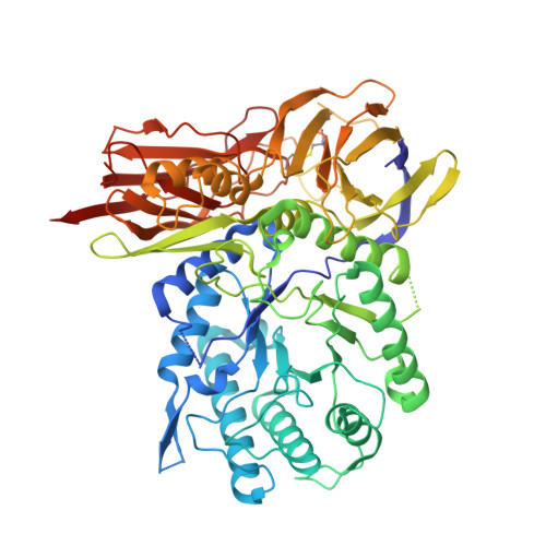
    6I6R, 6I6X

  • PubMed Abstract: 

    Cyclophellitol aziridines are potent irreversible inhibitors of retaining glycosidases and versatile intermediates in the synthesis of activity-based glycosidase probes (ABPs). Direct 3-amino-2-(trifluoromethyl)quinazolin-4(3H)-one-mediated aziridination of l-ido-configured cyclohexene has enabled the synthesis of new covalent inhibitors and ABPs of α-l-iduronidase, deficiency of which underlies the lysosomal storage disorder mucopolysaccharidosis type I (MPS I). The iduronidase ABPs react covalently and irreversibly in an activity-based manner with human recombinant α-l-iduronidase (rIDUA, Aldurazyme ® ). The structures of IDUA when complexed with the inhibitors in a non-covalent transition state mimicking form and a covalent enzyme-bound form provide insights into its conformational itinerary. Inhibitors 1-3 adopt a half-chair conformation in solution ( 4 H 3 and 3 H 4 ), as predicted by DFT calculations, which is different from the conformation of the Michaelis complex observed by crystallographic studies. Consequently, 1-3 may need to overcome an energy barrier in order to switch from the 4 H 3 conformation to the transition state ( 2, 5 B) binding conformation before reacting and adopting a covalent 5 S 1 conformation. rIDUA can be labeled with fluorescent Cy5 ABP 2, which allows monitoring of the delivery of therapeutic recombinant enzyme to lysosomes, as is intended in enzyme replacement therapy for the treatment of MPS I patients.


  • Organizational Affiliation
    • Department of Bio-organic Synthesis, Leiden Institute of Chemistry, Leiden University, Einsteinweg 55, 2333 CC, Leiden, The Netherlands.

Macromolecule Content 

  • Total Structure Weight: 143.04 kDa 
  • Atom Count: 9,957 
  • Modeled Residue Count: 1,196 
  • Deposited Residue Count: 1,254 
  • Unique protein chains: 1

Macromolecules

Find similar proteins by:|  3D Structure
Entity ID: 1
MoleculeChains  Sequence LengthOrganismDetailsImage
Alpha-L-iduronidase
A, B
627Homo sapiensMutation(s): 0 
EC: 3.2.1.76
UniProt & NIH Common Fund Data Resources
Find proteins for P35475 (Homo sapiens)
Explore P35475 
Go to UniProtKB:  P35475
PHAROS:  P35475
GTEx:  ENSG00000127415 
Entity Groups
Sequence Clusters30% Identity50% Identity70% Identity90% Identity95% Identity100% Identity
UniProt GroupP35475
Glycosylation
Glycosylation Sites: 3Go to GlyGen: P35475-1
Sequence Annotations
Expand
Reference Sequence

Oligosaccharides

Help  
Entity ID: 2
MoleculeChains Length2D Diagram GlycosylationD Interactions
beta-D-mannopyranose-(1-4)-2-acetamido-2-deoxy-beta-D-glucopyranose-(1-4)-2-acetamido-2-deoxy-beta-D-glucopyranose
C, D
3N-Glycosylation
Glycosylation Resources
GlyTouCan: G15407YE
GlyCosmos: G15407YE
GlyGen: G15407YE

Small Molecules

Ligands 5 Unique
IDChains Name / Formula / InChI Key2D Diagram3D Interactions
NAG

Query on NAG


Download:Ideal Coordinates CCD File
F [auth A],
G [auth A],
M [auth B],
N [auth B]
2-acetamido-2-deoxy-beta-D-glucopyranose
C8 H15 N O6
OVRNDRQMDRJTHS-FMDGEEDCSA-N
H62

Query on H62


Download:Ideal Coordinates CCD File
E [auth A],
L [auth B]
(1~{R},2~{R},3~{R},4~{S},6~{S})-6-azanyl-2,3,4-tris(oxidanyl)cyclohexane-1-carboxylic acid
C7 H13 N O5
DNFVBIRHFDBCAW-HGVZOGFYSA-N
TLA

Query on TLA


Download:Ideal Coordinates CCD File
J [auth A]L(+)-TARTARIC ACID
C4 H6 O6
FEWJPZIEWOKRBE-JCYAYHJZSA-N
GOL

Query on GOL


Download:Ideal Coordinates CCD File
H [auth A],
I [auth A],
O [auth B]
GLYCEROL
C3 H8 O3
PEDCQBHIVMGVHV-UHFFFAOYSA-N
CL

Query on CL


Download:Ideal Coordinates CCD File
K [auth A],
P [auth B]
CHLORIDE ION
Cl
VEXZGXHMUGYJMC-UHFFFAOYSA-M

Experimental Data & Validation

Experimental Data

  • Method: X-RAY DIFFRACTION
  • Resolution: 2.02 Å
  • R-Value Free:  0.240 (Depositor), 0.246 (DCC) 
  • R-Value Work:  0.213 (Depositor), 0.220 (DCC) 
  • R-Value Observed: 0.214 (Depositor) 
Space Group: P 21 21 21
Unit Cell:
Length ( Å )Angle ( ˚ )
a = 206.86α = 90
b = 69.89β = 90
c = 93.69γ = 90
Software Package:
Software NamePurpose
REFMACrefinement
xia2data reduction
xia2data scaling
PHASERphasing

Structure Validation

View Full Validation Report


Ligand Structure Quality Assessment 


Entry History 

Deposition Data

Revision History  (Full details and data files)

  • Version 1.0: 2018-12-05
    Type: Initial release
  • Version 1.1: 2018-12-26
    Changes: Data collection, Database references
  • Version 1.2: 2019-03-06
    Changes: Data collection
  • Version 1.3: 2019-08-14
    Changes: Data collection
  • Version 2.0: 2020-07-29
    Type: Remediation
    Reason: Carbohydrate remediation
    Changes: Advisory, Atomic model, Data collection, Derived calculations, Structure summary
  • Version 2.1: 2024-01-24
    Changes: Data collection, Database references, Refinement description, Structure summary
  • Version 2.2: 2024-11-13
    Changes: Structure summary