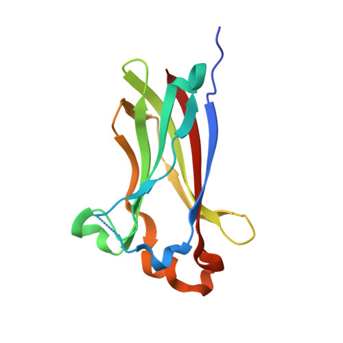
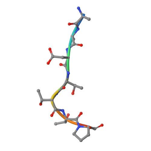
6I41

Co-crystal structure of human SPOP MATH domain (wild-type) and human BRD3 fragment


Experimental Data Snapshot

  • Method: X-RAY DIFFRACTION
  • Resolution: 1.90 Å
  • R-Value Free: 0.238 
  • R-Value Work: 0.188 
  • R-Value Observed: 0.190 

Starting Model: experimental
View more details

wwPDB Validation   3D Report Full Report


This is version 1.2 of the entry. See complete history


Literature

Structural Insights into BET Client Recognition of Endometrial and Prostate Cancer-Associated SPOP Mutants.

Ostertag, M.S.Hutwelker, W.Plettenburg, O.Sattler, M.Popowicz, G.M.

(2019) J Mol Biol 431: 2213-2221

  • DOI: https://doi.org/10.1016/j.jmb.2019.04.017
  • Primary Citation of Related Structures:  
    6I41, 6I5P, 6I68, 6I7A

  • PubMed Abstract: 

    BET proteins such as BRD3 are oncogenic transcriptional coactivators. SPOP binding triggers their proteasomal degradation. In both endometrial and prostate cancers, SPOP mutations occur in the MATH domain, but with opposed influence on drug susceptibility. In prostate cancer, SPOP mutations presumably cause increased BET levels, decreasing BET inhibitor drug susceptibility. As opposed, in endometrial cancer, decreased BET levels concomitant with higher BET inhibitor drug susceptibility were observed. Here, we present the to our knowledge first co-crystal structure of SPOP and a bromodomain containing protein (BRD3). Our structural and biophysical data confirm the suggested loss-of-function in prostate cancer-associated SPOP mutants and provide mechanistic explanation. As opposed to previous literature, our data on endometrial cancer-associated SPOP mutants do not show altered binding behavior compared to wild-type SPOP, indicating a more complex regulatory mechanism. SPOP mutation screening may thus be considered a valuable personalized medicine tool for effective antitumor therapy.


  • Organizational Affiliation

    Institute of Structural Biology, Helmholtz Zentrum München, Ingolstädter Landstr. 1, 85764 Neuherberg, Germany; Center for Integrated Protein Science Munich at Chair of Biomolecular NMR Spectroscopy, Department Chemie, Technische Universität München, Lichtenbergstrasse 4, 85747 Garching, Germany.


Macromolecules
Find similar proteins by:  (by identity cutoff)  |  3D Structure
Entity ID: 1
MoleculeChains Sequence LengthOrganismDetailsImage
Speckle-type POZ protein145Homo sapiensMutation(s): 0 
Gene Names: SPOP
UniProt & NIH Common Fund Data Resources
Find proteins for O43791 (Homo sapiens)
Explore O43791 
Go to UniProtKB:  O43791
PHAROS:  O43791
GTEx:  ENSG00000121067 
Entity Groups  
Sequence Clusters30% Identity50% Identity70% Identity90% Identity95% Identity100% Identity
UniProt GroupO43791
Sequence Annotations
Expand
  • Reference Sequence

Find similar proteins by:  Sequence   |   3D Structure  

Entity ID: 2
MoleculeChains Sequence LengthOrganismDetailsImage
Bromodomain-containing protein 39Homo sapiensMutation(s): 0 
Gene Names: BRD3KIAA0043RING3L
UniProt & NIH Common Fund Data Resources
Find proteins for Q15059 (Homo sapiens)
Explore Q15059 
Go to UniProtKB:  Q15059
PHAROS:  Q15059
GTEx:  ENSG00000169925 
Entity Groups  
UniProt GroupQ15059
Sequence Annotations
Expand
  • Reference Sequence
Experimental Data & Validation

Experimental Data

  • Method: X-RAY DIFFRACTION
  • Resolution: 1.90 Å
  • R-Value Free: 0.238 
  • R-Value Work: 0.188 
  • R-Value Observed: 0.190 
  • Space Group: C 1 2 1
Unit Cell:
Length ( Å )Angle ( ˚ )
a = 61.508α = 90
b = 55.18β = 100.39
c = 42.606γ = 90
Software Package:
Software NamePurpose
PHENIXrefinement
XDSdata reduction
XSCALEdata scaling
MOLREPphasing

Structure Validation

View Full Validation Report


Entry History 

Deposition Data

Revision History  (Full details and data files)

  • Version 1.0: 2019-05-08
    Type: Initial release
  • Version 1.1: 2019-06-05
    Changes: Data collection, Database references
  • Version 1.2: 2024-01-24
    Changes: Data collection, Database references, Refinement description