6I3L | pdb_00006i3l

Bilirubin oxidase from Myrothecium verrucaria, mutant W396F


Experimental Data Snapshot

  • Method: X-RAY DIFFRACTION
  • Resolution: 2.10 Å
  • R-Value Free: 
    0.196 (Depositor), 0.146 (DCC) 
  • R-Value Work: 
    0.159 (Depositor) 
  • R-Value Observed: 
    0.161 (Depositor) 

Starting Model: experimental
View more details

wwPDB Validation 3D Report Full Report

Validation slider image for 6I3L

This is version 2.2 of the entry. See complete history

Literature

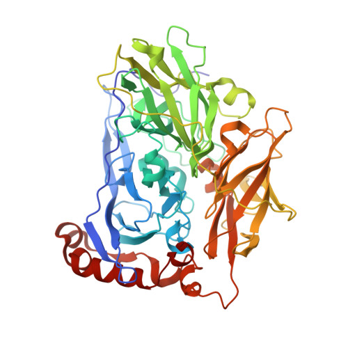
Trp-His covalent adduct in bilirubin oxidase is crucial for effective bilirubin binding but has a minor role in electron transfer.

Koval, T.Svecova, L.Ostergaard, L.H.Skalova, T.Duskova, J.Hasek, J.Kolenko, P.Fejfarova, K.Stransky, J.Trundova, M.Dohnalek, J.

(2019) Sci Rep 9: 13700-13700

  • DOI: https://doi.org/10.1038/s41598-019-50105-3
  • Primary Citation Related Structures: 
    6I3J, 6I3K, 6I3L

  • PubMed Abstract: 

    Unlike any protein studied so far, the active site of bilirubin oxidase from Myrothecium verrucaria contains a unique type of covalent link between tryptophan and histidine side chains. The role of this post-translational modification in substrate binding and oxidation is not sufficiently understood. Our structural and mutational studies provide evidence that this Trp396-His398 adduct modifies T1 copper coordination and is an important part of the substrate binding and oxidation site. The presence of the adduct is crucial for oxidation of substituted phenols and it substantially influences the rate of oxidation of bilirubin. Additionally, we bring the first structure of bilirubin oxidase in complex with one of its products, ferricyanide ion, interacting with the modified tryptophan side chain, Arg356 and the active site-forming loop 393-398. The results imply that structurally and chemically distinct types of substrates, including bilirubin, utilize the Trp-His adduct mainly for binding and to a smaller extent for electron transfer.


  • Organizational Affiliation
    • Institute of Biotechnology of the Czech Academy of Sciences, v.v.i., Průmyslová 595, 252 50, Vestec, Czech Republic. tomas.koval@ibt.cas.cz.

Macromolecule Content 

  • Total Structure Weight: 123.86 kDa 
  • Atom Count: 10,164 
  • Modeled Residue Count: 1,068 
  • Deposited Residue Count: 1,068 
  • Unique protein chains: 1

Macromolecules

Find similar proteins by:|  3D Structure
Entity ID: 1
MoleculeChains  Sequence LengthOrganismDetailsImage
Bilirubin oxidase
A, B
534Albifimbria verrucariaMutation(s): 1 
EC: 1.3.3.5
UniProt
Find proteins for Q12737 (Albifimbria verrucaria)
Explore Q12737 
Go to UniProtKB:  Q12737
Entity Groups
Sequence Clusters30% Identity50% Identity70% Identity90% Identity95% Identity100% Identity
UniProt GroupQ12737
Glycosylation
Glycosylation Sites: 2
Sequence Annotations
Expand
Reference Sequence

Oligosaccharides

Help  
Entity ID: 2
MoleculeChains Length2D Diagram GlycosylationD Interactions
beta-D-mannopyranose-(1-4)-2-acetamido-2-deoxy-beta-D-glucopyranose-(1-4)-2-acetamido-2-deoxy-beta-D-glucopyranose
C, E
3N-Glycosylation
Glycosylation Resources
GlyTouCan: G15407YE
GlyCosmos: G15407YE
GlyGen: G15407YE
Entity ID: 3
MoleculeChains Length2D Diagram GlycosylationD Interactions
2-acetamido-2-deoxy-beta-D-glucopyranose-(1-4)-2-acetamido-2-deoxy-beta-D-glucopyranose
D, F
2N-Glycosylation
Glycosylation Resources
GlyTouCan: G42666HT
GlyCosmos: G42666HT
GlyGen: G42666HT

Small Molecules

Ligands 5 Unique
IDChains Name / Formula / InChI Key2D Diagram3D Interactions
SIN

Query on SIN


Download:Ideal Coordinates CCD File
K [auth A],
L [auth A],
W [auth B],
X [auth B]
SUCCINIC ACID
C4 H6 O4
KDYFGRWQOYBRFD-UHFFFAOYSA-N
GOL

Query on GOL


Download:Ideal Coordinates CCD File
AA [auth B]
BA [auth B]
CA [auth B]
M [auth A]
N [auth A]
AA [auth B],
BA [auth B],
CA [auth B],
M [auth A],
N [auth A],
O [auth A],
P [auth A],
Q [auth A],
Z [auth B]
GLYCEROL
C3 H8 O3
PEDCQBHIVMGVHV-UHFFFAOYSA-N
CU

Query on CU


Download:Ideal Coordinates CCD File
G [auth A]
H [auth A]
I [auth A]
J [auth A]
S [auth B]
G [auth A],
H [auth A],
I [auth A],
J [auth A],
S [auth B],
T [auth B],
U [auth B],
V [auth B]
COPPER (II) ION
Cu
JPVYNHNXODAKFH-UHFFFAOYSA-N
OXY

Query on OXY


Download:Ideal Coordinates CCD File
DA [auth B],
R [auth A]
OXYGEN MOLECULE
O2
MYMOFIZGZYHOMD-UHFFFAOYSA-N
NA

Query on NA


Download:Ideal Coordinates CCD File
Y [auth B]SODIUM ION
Na
FKNQFGJONOIPTF-UHFFFAOYSA-N

Experimental Data & Validation

Experimental Data

  • Method: X-RAY DIFFRACTION
  • Resolution: 2.10 Å
  • R-Value Free:  0.196 (Depositor), 0.146 (DCC) 
  • R-Value Work:  0.159 (Depositor) 
  • R-Value Observed: 0.161 (Depositor) 
Space Group: F 2 2 2
Unit Cell:
Length ( Å )Angle ( ˚ )
a = 136.25α = 90
b = 200.663β = 90
c = 217.121γ = 90
Software Package:
Software NamePurpose
REFMACrefinement
XDSdata reduction
XDSdata scaling
MOLREPphasing

Structure Validation

View Full Validation Report


Entry History 

& Funding Information

Deposition Data


Funding OrganizationLocationGrant Number
Ministry of Education, Youth and Sports of the Czech RepublicCzech RepublicLM2015043
European Regional Development FundCzech RepublicCZ.02.1.01/0.0/0.0/15_003/0000447
European Regional Development FundCzech RepublicCZ.02.1.01/0.0/0.0/16_013/0001776
European Regional Development FundCzech RepublicCZ.1.05/1.1.00/02.0109
Czech Academy of SciencesCzech Republic86652036

Revision History  (Full details and data files)

  • Version 1.0: 2019-10-02
    Type: Initial release
  • Version 2.0: 2020-07-29
    Type: Remediation
    Reason: Carbohydrate remediation
    Changes: Atomic model, Data collection, Derived calculations, Structure summary
  • Version 2.1: 2024-05-01
    Changes: Data collection, Database references, Refinement description, Structure summary
  • Version 2.2: 2024-10-23
    Changes: Structure summary