


p53 dynamics vary between tissues and are linked with radiation sensitivity
Ornstein, J., Iwamoto, Y., Prytyskach, M., Miller, M., Kallen, J., Ferretti, S., Holzer, P., Forrester, W., Weissleder, R., Lahav, G.To be published.
Experimental Data Snapshot
Starting Model: experimental
View more details
Entity ID: 1 | |||||
|---|---|---|---|---|---|
| Molecule | Chains | Sequence Length | Organism | Details | Image |
| Human E3 Ubiquitin-Protein Ligase MDM2 | 96 | Homo sapiens | Mutation(s): 0 EC: 2.3.2.27 | 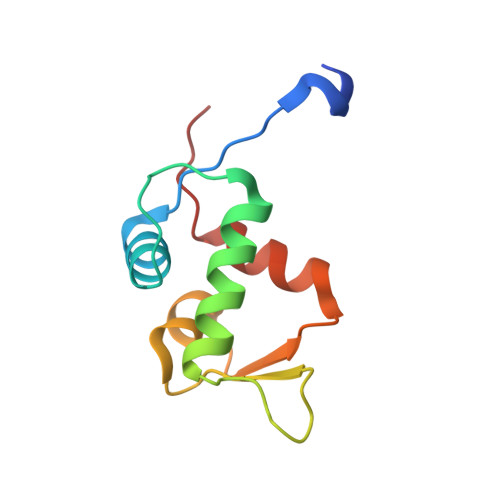 | |
UniProt & NIH Common Fund Data Resources | |||||
PHAROS: Q00987 GTEx: ENSG00000135679 | |||||
Entity Groups | |||||
| Sequence Clusters | 30% Identity50% Identity70% Identity90% Identity95% Identity100% Identity | ||||
| UniProt Group | Q00987 | ||||
Sequence AnnotationsExpand | |||||
Reference Sequence | |||||
| Ligands 1 Unique | |||||
|---|---|---|---|---|---|
| ID | Chains | Name / Formula / InChI Key | 2D Diagram | 3D Interactions | |
| H0W (Subject of Investigation/LOI) Download:Ideal Coordinates CCD File | B [auth A] | 6-chloranyl-3-[3-[(1~{S})-1-(4-chlorophenyl)ethyl]-5-phenyl-imidazol-4-yl]-~{N}-[2-[4-(2-oxidanylidene-1,3-oxazinan-3-yl)piperidin-1-yl]pyridin-3-yl]-1~{H}-indole-2-carboxamide C40 H37 Cl2 N7 O3 PEGKHNWZGSKUJH-VWLOTQADSA-N |  | ||
| Length ( Å ) | Angle ( ˚ ) |
|---|---|
| a = 121.883 | α = 90 |
| b = 121.883 | β = 90 |
| c = 121.883 | γ = 90 |
| Software Name | Purpose |
|---|---|
| XDS | data reduction |
| XSCALE | data scaling |
| PHASER | phasing |
| REFMAC | refinement |
| PDB_EXTRACT | data extraction |


