


Apo structure of TP domain from Chlamydia trachomatis penicillin-binding protein 3
Bellini, D., Koekemoer, L., Newman, H., Dowson, C.G.To be published.
Experimental Data Snapshot
Starting Model: experimental
View more details
wwPDB Validation 3D Report Full Report

Entity ID: 1 | |||||
|---|---|---|---|---|---|
| Molecule | Chains | Sequence Length | Organism | Details | Image |
| Penicillin-binding protein | 346 | Chlamydia trachomatis | Mutation(s): 2 Gene Names: pbp_2, ERS133248_00492 EC: 2.4.1.129 | 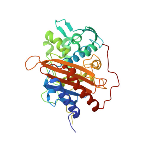 | |
| Length ( Å ) | Angle ( ˚ ) |
|---|---|
| a = 105.444 | α = 90 |
| b = 105.444 | β = 90 |
| c = 118.522 | γ = 120 |
| Software Name | Purpose |
|---|---|
| REFMAC | refinement |
| xia2 | data reduction |
| Aimless | data scaling |
| MrBUMP | phasing |
| Funding Organization | Location | Grant Number |
|---|---|---|
| Medical Research Council (United Kingdom) | United Kingdom | grant.MR/P007503/1 |


