6HWM | pdb_00006hwm

Structure of Thermus thermophilus ClpP in complex with bortezomib


Experimental Data Snapshot

  • Method: X-RAY DIFFRACTION
  • Resolution: 2.70 Å
  • R-Value Free: 
    0.236 (Depositor), 0.238 (DCC) 
  • R-Value Work: 
    0.205 (Depositor), 0.205 (DCC) 
  • R-Value Observed: 
    0.208 (Depositor) 

Starting Model: experimental
View more details

wwPDB Validation 3D Report Full Report

Validation slider image for 6HWM

Ligand Structure Quality Assessment 


This is version 1.4 of the entry. See complete history

Literature

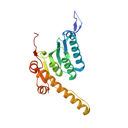
Mechanism of the allosteric activation of the ClpP protease machinery by substrates and active-site inhibitors.

Felix, J.Weinhaupl, K.Chipot, C.Dehez, F.Hessel, A.Gauto, D.F.Morlot, C.Abian, O.Gutsche, I.Velazquez-Campoy, A.Schanda, P.Fraga, H.

(2019) Sci Adv 5: eaaw3818-eaaw3818

  • DOI: https://doi.org/10.1126/sciadv.aaw3818
  • Primary Citation Related Structures: 
    6HWM, 6HWN

  • PubMed Abstract: 

    Coordinated conformational transitions in oligomeric enzymatic complexes modulate function in response to substrates and play a crucial role in enzyme inhibition and activation. Caseinolytic protease (ClpP) is a tetradecameric complex, which has emerged as a drug target against multiple pathogenic bacteria. Activation of different ClpPs by inhibitors has been independently reported from drug development efforts, but no rationale for inhibitor-induced activation has been hitherto proposed. Using an integrated approach that includes x-ray crystallography, solid- and solution-state nuclear magnetic resonance, molecular dynamics simulations, and isothermal titration calorimetry, we show that the proteasome inhibitor bortezomib binds to the ClpP active-site serine, mimicking a peptide substrate, and induces a concerted allosteric activation of the complex. The bortezomib-activated conformation also exhibits a higher affinity for its cognate unfoldase ClpX. We propose a universal allosteric mechanism, where substrate binding to a single subunit locks ClpP into an active conformation optimized for chaperone association and protein processive degradation.


  • Organizational Affiliation
    • Institut de Biologie Structurale, Université Grenoble Alpes, CEA, CNRS, IBS, 71 Avenue des Martyrs, F-38044 Grenoble, France.

Macromolecule Content 

  • Total Structure Weight: 162.35 kDa 
  • Atom Count: 10,130 
  • Modeled Residue Count: 1,311 
  • Deposited Residue Count: 1,428 
  • Unique protein chains: 1

Macromolecules

Find similar proteins by:|  3D Structure
Entity ID: 1
MoleculeChains  Sequence LengthOrganismDetailsImage
ATP-dependent Clp protease proteolytic subunit
A, B, C, D, E
A, B, C, D, E, F, G
204Thermus thermophilusMutation(s): 0 
Gene Names: clpPTTHA0615
EC: 3.4.21.92
UniProt
Find proteins for Q5SKM8 (Thermus thermophilus (strain ATCC 27634 / DSM 579 / HB8))
Explore Q5SKM8 
Go to UniProtKB:  Q5SKM8
Entity Groups
Sequence Clusters30% Identity50% Identity70% Identity90% Identity95% Identity100% Identity
UniProt GroupQ5SKM8
Sequence Annotations
Expand
Reference Sequence

Small Molecules

Ligands 2 Unique
IDChains Name / Formula / InChI Key2D Diagram3D Interactions
BO2

Query on BO2


Download:Ideal Coordinates CCD File
H [auth A]
I [auth B]
J [auth C]
L [auth D]
M [auth E]
H [auth A],
I [auth B],
J [auth C],
L [auth D],
M [auth E],
O [auth F],
P [auth G]
N-[(1R)-1-(DIHYDROXYBORYL)-3-METHYLBUTYL]-N-(PYRAZIN-2-YLCARBONYL)-L-PHENYLALANINAMIDE
C19 H25 B N4 O4
GXJABQQUPOEUTA-RDJZCZTQSA-N
PEG

Query on PEG


Download:Ideal Coordinates CCD File
K [auth C],
N [auth E],
Q [auth G]
DI(HYDROXYETHYL)ETHER
C4 H10 O3
MTHSVFCYNBDYFN-UHFFFAOYSA-N

Experimental Data & Validation

Experimental Data

  • Method: X-RAY DIFFRACTION
  • Resolution: 2.70 Å
  • R-Value Free:  0.236 (Depositor), 0.238 (DCC) 
  • R-Value Work:  0.205 (Depositor), 0.205 (DCC) 
  • R-Value Observed: 0.208 (Depositor) 
Space Group: C 2 2 21
Unit Cell:
Length ( Å )Angle ( ˚ )
a = 135.139α = 90
b = 168.74β = 90
c = 166.083γ = 90
Software Package:
Software NamePurpose
PHENIXrefinement
XDSdata reduction
XDSdata scaling
PHASERphasing

Structure Validation

View Full Validation Report


Ligand Structure Quality Assessment 


Entry History 

& Funding Information

Deposition Data


Funding OrganizationLocationGrant Number
Grenoble Instruct-ERIC CenterFrance301
Grenoble Alliance for Integrated Structural Cell BiologyFranceANR-10-LABX-49-01
French Infrastructure for Integrated Structural BiologyFranceANR-10-INSB-05-02

Revision History  (Full details and data files)

  • Version 1.0: 2019-09-18
    Type: Initial release
  • Version 1.1: 2019-09-25
    Changes: Data collection, Database references
  • Version 1.2: 2023-04-12
    Changes: Database references, Source and taxonomy
  • Version 1.3: 2024-02-07
    Changes: Data collection, Refinement description
  • Version 1.4: 2024-10-09
    Changes: Structure summary