


Human SLK bound to a maleimide inhibitor
Sorrell, F.J., Berger, B.T., Salah, E., Serafim, R.A.M., Elkins, J.M.To be published.
Experimental Data Snapshot
Starting Model: experimental
View more details
Entity ID: 1 | |||||
|---|---|---|---|---|---|
| Molecule | Chains | Sequence Length | Organism | Details | Image |
| STE20-like serine/threonine-protein kinase | 304 | Homo sapiens | Mutation(s): 0 Gene Names: SLK, KIAA0204, STK2 EC: 2.7.11.1 | 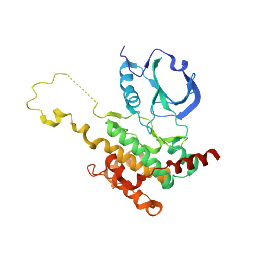 | |
UniProt & NIH Common Fund Data Resources | |||||
PHAROS: Q9H2G2 GTEx: ENSG00000065613 | |||||
Entity Groups | |||||
| Sequence Clusters | 30% Identity50% Identity70% Identity90% Identity95% Identity100% Identity | ||||
| UniProt Group | Q9H2G2 | ||||
Sequence AnnotationsExpand | |||||
Reference Sequence | |||||
| Ligands 3 Unique | |||||
|---|---|---|---|---|---|
| ID | Chains | Name / Formula / InChI Key | 2D Diagram | 3D Interactions | |
| GUQ (Subject of Investigation/LOI) Download:Ideal Coordinates CCD File | E [auth A] | 3-(1,3-benzothiazol-2-ylamino)-4-(2-methoxyphenyl)pyrrole-2,5-dione C18 H13 N3 O3 S DKODQPUAYQCESR-UHFFFAOYSA-N |  | ||
| SO4 Download:Ideal Coordinates CCD File | F [auth A], G [auth A] | SULFATE ION O4 S QAOWNCQODCNURD-UHFFFAOYSA-L |  | ||
| EDO Download:Ideal Coordinates CCD File | B [auth A], C [auth A], D [auth A] | 1,2-ETHANEDIOL C2 H6 O2 LYCAIKOWRPUZTN-UHFFFAOYSA-N |  | ||
| Length ( Å ) | Angle ( ˚ ) |
|---|---|
| a = 69.871 | α = 90 |
| b = 69.871 | β = 90 |
| c = 164.301 | γ = 90 |
| Software Name | Purpose |
|---|---|
| XDS | data reduction |
| Aimless | data scaling |
| PHASER | phasing |
| PHENIX | refinement |
| PDB_EXTRACT | data extraction |


